. 2008 Nov;190(22):7392-405.
doi: 10.1128/JB.00564-08. Epub 2008 Sep 12.

Identification and characterization of cyclic diguanylate signaling systems controlling rugosity in Vibrio cholerae

Affiliations

Identification and characterization of cyclic diguanylate signaling systems controlling rugosity in Vibrio cholerae

Sinem Beyhan et al. J Bacteriol. 2008 Nov.

Abstract

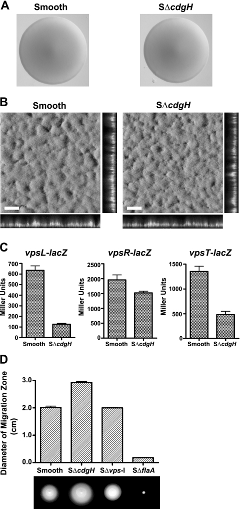
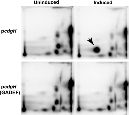
Vibrio cholerae, the causative agent of the disease cholera, can generate rugose variants that have an increased capacity to form biofilms. Rugosity and biofilm formation are critical for the environmental survival and transmission of the pathogen, and these processes are controlled by cyclic diguanylate (c-di-GMP) signaling systems. c-di-GMP is produced by diguanylate cyclases (DGCs) and degraded by phosphodiesterases (PDEs). Proteins that contain GGDEF domains act as DGCs, whereas proteins that contain EAL or HD-GYP domains act as PDEs. In the V. cholerae genome there are 62 genes that are predicted to encode proteins capable of modulating the cellular c-di-GMP concentration. We previously identified two DGCs, VpvC and CdgA, that can control the switch between smooth and rugose. To identify other c-di-GMP signaling proteins involved in rugosity, we generated in-frame deletion mutants of all genes predicted to encode proteins with GGDEF and EAL domains and then searched for mutants with altered rugosity. In this study, we identified two new genes, cdgG and cdgH, involved in rugosity control. We determined that CdgH acts as a DGC and positively regulates rugosity, whereas CdgG does not have DGC activity and negatively regulates rugosity. In addition, epistasis analysis with CdgG, CdgH, and other DGCs and PDEs controlling rugosity revealed that CdgG and CdgH act in parallel with previously identified c-di-GMP signaling proteins to control rugosity in V. cholerae. We also determined that PilZ domain-containing c-di-GMP binding proteins contribute minimally to rugosity, indicating that there are additional c-di-GMP binding proteins controlling rugosity in V. cholerae.


Figures

FIG. 1.
FIG. 1.
Phenotypic characterization of CdgH in the rugose genetic background. (A) Colony morphologies of rugose and RΔcdgH strains that were grown for 24 and 48 h on LB agar plates at 30°C. (B) Three-dimensional biofilm structures of rugose and RΔcdgH strains that are formed 8 h postinoculation under static conditions at 30°C. Images were acquired with CLSM, with top-down (large panes) and orthogonal (side panels) views of biofilms shown. Scale bar, 30 μm. (C) Transcription of vpsL-lacZ, vpsR-lacZ, and vpsT-lacZ fusions determined in rugose and RΔcdgH strains by measuring β-galactosidase activity in cells that were grown to mid-exponential phase in LB medium at 30°C. The result shown is representative of three independent experiments. Error bars represent the standard deviations. (D) Diameter of migration zone of rugose, RΔcdgH, RΔvps-I, and RΔflaA strains measured in LB soft agar plates (0.3%) after 18 h of incubation at 30°C. The data shown are representative of three independent experiments. Error bars represent the standard deviations.
FIG. 2.
FIG. 2.
Phenotypic characterization of CdgH in the smooth genetic background. (A) Colony morphologies of smooth and SΔcdgH strains that were grown for 24 h on LB agar plates at 30°C. (B) Three-dimensional biofilm structures of smooth and SΔcdgH strains that are formed 24 h postinoculation in a once-through flow cell system. Images were acquired with CLSM, with top-down (large panes) and orthogonal (side panels) views of biofilms shown. Scale bar, 30 μm. (C) Transcription of vpsL-lacZ, vpsR-lacZ, and vpsT-lacZ fusions determined in smooth and SΔcdgH strains by measuring β-galactosidase activity in cells that were grown to mid-exponential phase in LB medium at 30°C. The result shown is representative of three independent experiments. Error bars represent the standard deviations. (D) Diameter of migration zone of smooth, SΔcdgH, SΔvps-I, and SΔflaA strains measured in LB soft agar plates (0.3%) after 18 h of incubation at 30°C. The data shown are representative of three independent experiments. Error bars represent the standard deviations.
FIG. 3.
FIG. 3.
Analysis of enzymatic activity of CdgH. 2D-TLC analysis of total nucleotides extracted from smooth strains harboring pcdgH or pcdgH(GADEF) grown in MOPS minimal medium with [32P]orthophosphate was performed. The arrow indicates the spot corresponding to c-di-GMP according to its Rf values of 0.16 in the NH4CO3 dimension and 0.37 in the KH2PO4 dimension. The result shown are representative of two independent experiments.
FIG. 4.
FIG. 4.
Phenotypic characterization of CdgG in the rugose genetic background. (A) Colony morphologies of rugose and RΔcdgG strains that were grown for 24 and 48 h on LB agar plates at 30°C. (B) Three-dimensional biofilm structures of rugose and RΔcdgG strains that are formed 8 h postinoculation under static conditions at 30°C. Images were acquired with CLSM, with top-down (large panes) and orthogonal (side panels) views of biofilms shown. Scale bar, 30 μm. (C) Transcription of vpsL-lacZ, vpsR-lacZ, and vpsT-lacZ fusions determined in rugose and RΔcdgG strains by measuring β-galactosidase activity in cells that were grown to mid-exponential phase in LB medium at 30°C. The results shown are representative of three independent experiments. Error bars represent the standard deviations. (D) Diameter of migration zone of rugose, RΔcdgG, RΔvps-I, and RΔflaA strains measured in LB soft agar plates (0.3%) after 18 h of incubation at 30°C. The data shown are representative of three independent experiments. Error bars represent the standard deviations.
FIG. 5.
FIG. 5.
Phenotypic characterization of CdgG in the smooth genetic background. (A) Colony morphologies of smooth and SΔcdgG strains that were grown for 24 h on LB agar plates at 30°C. (B) Three-dimensional biofilm structures of smooth and SΔcdgG strains that are formed 24 h postinoculation in a once-through flow cell system. Images were acquired with CLSM, with top-down (large panes) and orthogonal (side panels) views of biofilms shown. Scale bar, 30 μm. (C) Transcription of vpsL-lacZ, vpsR-lacZ, and vpsT-lacZ fusions determined in smooth and SΔcdgG strains by measuring β-galactosidase activity in cells that were grown to mid-exponential phase in LB medium at 30°C. The results shown are representative of three independent experiments. Error bars represent the standard deviations. (D) Diameter of migration zone of smooth, SΔcdgG, SΔvps-I, and SΔflaA strains measured in LB soft agar plates (0.3%) after 18 h of incubation at 30°C. The data shown are representative of three independent experiments. Error bars represent the standard deviations.
FIG. 6.
FIG. 6.
Analysis of enzymatic activity of CdgG. 2D-TLC analysis of total nucleotides extracted from smooth strains harboring pcdgG or pcdgG(SGAAF) grown in MOPS minimal medium with [32P]orthophosphate was performed. The results shown are representative of two independent experiments.
FIG. 7.
FIG. 7.
Epistasis analysis of cdgG and cdgH. (A) Colony morphologies of rugose, RΔcdgG, RΔcdgH, RΔcdgGΔcdgH, smooth, SΔcdgG, SΔcdgH, and SΔcdgGΔcdgH strains that were grown for 24 h on LB agar plates at 30°C. (B) Diameter of migration zone of rugose, RΔcdgG, RΔcdgH, RΔcdgGΔcdgH, smooth, SΔcdgG, SΔcdgH, and SΔcdgGΔcdgH strains measured in LB soft agar plates (0.3%) after 18 h of incubation at 30°C. The data shown are representative of three independent experiments. Error bars represent the standard deviations.
FIG. 8.
FIG. 8.
Epistasis analysis of cdgH, vpvC, and cdgA. (A) Colony morphologies of rugose, RΔcdgH, RΔvpvC, and RΔvpvCΔcdgH strains that were grown for 24 h on LB-agar plates at 30°C. (B) Diameter of migration zone of rugose, RΔcdgH, RΔvpvC, and RΔvpvCΔcdgH strains measured in LB soft agar plates (0.3%) after 18 h of incubation at 30°C. The data shown are representative of three independent experiments. Error bars represent the standard deviations. (C) Colony morphologies of smooth, SΔcdgA, SΔcdgH, SΔcdgAΔcdgH, SΔhapR, SΔhapRΔcdgA, SΔhapRΔcdgH, and SΔhapRΔcdgAΔcdgH that were grown for 48 h on LB agar plates at 30°C.
FIG. 9.
FIG. 9.
Epistasis analysis of rocS, mbaA, cdgC and cdgG in the smooth genetic background. Colony morphologies of smooth, SΔrocS, SΔmbaA, SΔcdgC, SΔrocΔmbaAΔcdgC, SΔcdgG, SΔrocSΔcdgG, SΔmbaAΔcdgG, SΔcdgCΔcdgG, and SΔrocSΔmbaAΔcdgCΔcdgG strains that were grown for 48 h on LB agar plates at 30°C.
FIG. 10.
FIG. 10.
Characterization plz genes for their contribution to rugosity. (A) Colony morphologies of rugose, RΔplzA, RΔplzB, RΔplzC, RΔplzD, RΔplzE, RΔplzACDE, and RΔplzABCDE strains that were grown for 48 h on LB agar plates at 30°C. (B) Three-dimensional biofilm structures of rugose and RΔplzACDE strains that are formed at 24 h postinoculation in a once-through flow cell system. Images were acquired with CLSM, with top-down (large panes) and orthogonal (side panels) views of biofilms shown. The white bar equals 30 μm. (C) Colony morphologies of rugose, RΔcdgH, RΔvpvC, RΔplzC, RΔcdgHΔplzC, and RΔvpvCΔplzC strains that were grown for 24 h on LB agar plates at 30°C. (D) Diameter of migration zone of rugose, RΔplzC, RΔcdgH, RΔcdgHΔplzC, RΔvpvC, and RΔvpvCΔplzC strains measured in LB soft agar plates (0.3%) after 18 h of incubation at 30°C. The data shown are representative of three independent experiments. Error bars represent the standard deviations.
FIG. 11.
FIG. 11.
Characterization of A-site and I-site of CdgG. (A) Multiple sequence alignment of C. crescentus protein PleD and CdgG at the predicted I-site and A-site of GGDEF domain is shown. Conserved residues are highlighted, and the marked residues (Arg434, Asp437, Glu445, and Glu446) were changed to alanine residues via site-directed mutagenesis. (B) Colony morphologies of RΔcdgG strains harboring pBAD/myc-His, pcdgG, pcdgG(SGAAF), or pcdgG(ADSA) that were grown for 24 h at 30°C on LB agar plates containing 0.02% (wt/vol) l-arabinose. (C) Three-dimensional biofilm structures of SΔcdgG strains harboring pBAD/myc-His, pcdgG, pcdgG(SGAAF), or pcdgG(ADSA) that are formed 24 h postinoculation in a once-through flow cell system. Images were acquired with CLSM, with top-down (large panes) and orthogonal (side panels) views of biofilms shown. Scale bar, 30 μm.
FIG. 12.
FIG. 12.
A model of c-di-GMP signaling systems modulating rugosity-associated phenotypes in V. cholerae. VpvC, CdgA, and CdgH are localized in the cytoplasmic membrane, generating different pools of c-di-GMP upon receiving environmental cues. Intracellular c-di-GMP is degraded by membrane localized MbaA and cytoplasmically localized RocS and CdgC. The function of CdgG in the system is unclear, although it is likely to interact with RocS. CdgG can act as a c-di-GMP binding protein. Through Plz proteins and other c-di-GMP binding proteins and then, via effector proteins, c-di-GMP signaling systems affect colony corrugation, biofilm formation, VPS production, and motility in V. cholerae.

References

    1. Alam, M., M. Sultana, G. B. Nair, R. B. Sack, D. A. Sack, A. K. Siddique, A. Ali, A. Huq, and R. R. Colwell. 2006. Toxigenic Vibrio cholerae in the aquatic environment of Mathbaria, Bangladesh. Appl. Environ. Microbiol. 722849-2855. - PMC - PubMed
    1. Amikam, D., and M. Y. Galperin. 2006. PilZ domain is part of the bacterial c-di-GMP binding protein. Bioinformatics 223-6. - PubMed
    1. Bao, Y., D. P. Lies, H. Fu, and G. P. Roberts. 1991. An improved Tn7-based system for the single-copy insertion of cloned genes into chromosomes of gram-negative bacteria. Gene 109167-168. - PubMed
    1. Beyhan, S., K. Bilecen, S. R. Salama, C. Casper-Lindley, and F. H. Yildiz. 2007. Regulation of rugosity and biofilm formation in Vibrio cholerae: comparison of VpsT and VpsR regulons and epistasis analysis of vpsT, vpsR, and hapR. J. Bacteriol. 189388-402. - PMC - PubMed
    1. Beyhan, S., A. D. Tischler, A. Camilli, and F. H. Yildiz. 2006. Transcriptome and phenotypic responses of Vibrio cholerae to increased cyclic di-GMP level. J. Bacteriol. 1883600-3613. - PMC - PubMed

Publication types

MeSH terms

LinkOut - more resources