

Three-dimensional surface mapping of the caudate nucleus in late-life depression
- PMID: 18790876
- PMCID: PMC2970509
- DOI: 10.1097/JGP.0b013e31816ff72b
Three-dimensional surface mapping of the caudate nucleus in late-life depression
Abstract
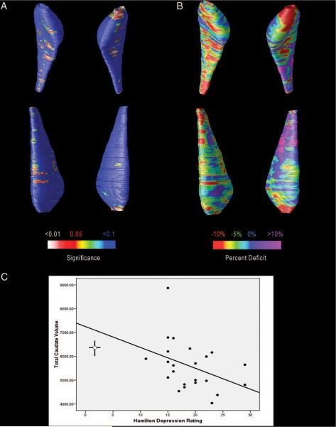
Objective: To compare the volumes of the caudate nucleus, using traditional volumetry and a three-dimensional brain mapping technique, in a group of individuals with late-life depression and a group of age- and education-equated nondepressed comparison subjects.
Design: Cross-sectional.
Setting: University Medical Center.
Participants: Twenty-three nondemented subjects with late-life depression and 15 age- and education-equated elderly comparison subjects (depressed mean years of age: 70.5 +/- 5.7 SD, comparison subjects = 69.9 years +/- 6.4) with no history of psychiatric or neurologic disease.
Measurements: Structural magnetic resonance imaging. Three-dimensional (3-D) surface models were created from manually traced outlines of the caudate nucleus from spoiled gradient echo images. Models were geometrically averaged across subjects and statistical maps created to localize any regional volume differences between groups.
Results: Relative to comparison subjects, depressed subjects had significantly lower mean volumes for both the left (p = 0.029) and right (p = 0.052) caudate nucleus as well as total caudate volume (p = 0.032). Total volumes were 13.1% less in the depressed group (13.5% on the left and 12.6% on the right). 3-D maps further localized these reductions to the caudate head. Volume reductions were correlated with depression severity, as measured by the 17-item Hamilton Depression Rating Scale.
Conclusion: Late-life depression is associated with left and right caudate nucleus reduction especially in anterior portions. Among depressed subjects, greater caudate reduction was associated with more severe depression. These results are consistent with growing evidence that the anterior caudate nucleus, especially the head, may be structurally and functionally abnormal in affective disorders.
Figures
References
-
- Morris P, Rapoport SI. Neuroimaging and affective disorder in late life: a review. Can J Psychiatry. 1990;35:347–354. - PubMed
-
- Coffey CE, Figiel GS, Djang WT, et al. Subcortical hyperintensity on magnetic resonance imaging: a comparison of normal and depressed elderly subjects. Am J Psychiatry. 1990;147:187–189. - PubMed
-
- Hickie I, Scott E, Mitchell P, et al. Subcortical hyperintensities on magnetic resonance imaging: clinical correlates and prognostic significance in patients with severe depression. Biol Psychiatry. 1995;37:151–160. - PubMed
-
- Krishnan KR, McDonald WM, Doraiswamy PM, et al. Neuroanatomical substrates of depression in the elderly. Eur Arch Psychiatry Clin Neurosci. 1993;243:41–46. - PubMed
-
- Lesser IM, Boone KB, Mehringer CM, et al. Cognition and white matter hyperintensities in older depressed patients. Am J Psychiatry. 1996;153:1280–1287. - PubMed
Publication types
MeSH terms
Grants and funding
- MH 71955/MH/NIMH NIH HHS/United States
- AG 05133/AG/NIA NIH HHS/United States
- RR 019771/RR/NCRR NIH HHS/United States
- K01 MH001684/MH/NIMH NIH HHS/United States
- MH 52247/MH/NIMH NIH HHS/United States
- P30 MH071944/MH/NIMH NIH HHS/United States
- P50 AG016570/AG/NIA NIH HHS/United States
- R01 MH072947/MH/NIMH NIH HHS/United States
- MH 072947/MH/NIMH NIH HHS/United States
- AG 016570/AG/NIA NIH HHS/United States
- HD 050735/HD/NICHD NIH HHS/United States
- P30 MH052247/MH/NIMH NIH HHS/United States
- P30 AG024827/AG/NIA NIH HHS/United States
- R21 RR019771/RR/NCRR NIH HHS/United States
- MH 071944/MH/NIMH NIH HHS/United States
- R01 HD050735/HD/NICHD NIH HHS/United States
- MH 01684/MH/NIMH NIH HHS/United States
- EB 01651/EB/NIBIB NIH HHS/United States
- P50 AG005133/AG/NIA NIH HHS/United States
- P41 RR013642/RR/NCRR NIH HHS/United States
LinkOut - more resources
Full Text Sources
Medical
