Clinical Trial
. 2008 Sep;248(3):387-401.
doi: 10.1097/SLA.0b013e3181856241.

Pathophysiologic response to severe burn injury

Affiliations
Clinical Trial

Pathophysiologic response to severe burn injury

Marc G Jeschke et al. Ann Surg. 2008 Sep.

Abstract

Objective: To improve clinical outcome and to determine new treatment options, we studied the pathophysiologic response postburn in a large prospective, single center, clinical trial.

Summary background data: A severe burn injury leads to marked hypermetabolism and catabolism, which are associated with morbidity and mortality. The underlying pathophysiology and the correlations between humoral changes and organ function have not been well delineated.

Methods: Two hundred forty-two severely burned pediatric patients [>30% total body surface area (TBSA)], who received no anabolic drugs, were enrolled in this study. Demographics, clinical data, serum hormones, serum cytokine expression profile, organ function, hypermetabolism, muscle protein synthesis, incidence of wound infection sepsis, and body composition were obtained throughout acute hospital course.

Results: Average age was 8 +/- 0.2 years, and average burn size was 56 +/- 1% TBSA with 43 +/- 1% third-degree TBSA. All patients were markedly hypermetabolic throughout acute hospital stay and had significant muscle protein loss as demonstrated by a negative muscle protein net balance (-0.05% +/- 0.007 nmol/100 mL leg/min) and loss of lean body mass (LBM) (-4.1% +/- 1.9%); P < 0.05. Patients lost 3% +/- 1% of their bone mineral content (BMC) and 2 +/- 1% of their bone mineral density (BMD). Serum proteome analysis demonstrated profound alterations immediately postburn, which remained abnormal throughout acute hospital stay; P < 0.05. Cardiac function was compromised immediately after burn and remained abnormal up to discharge; P < 0.05. Insulin resistance appeared during the first week postburn and persisted until discharge. Patients were hyperinflammatory with marked changes in IL-8, MCP-1, and IL-6, which were associated with 2.5 +/- 0.2 infections and 17% sepsis.

Conclusions: In this large prospective clinical trial, we delineated the complexity of the postburn pathophysiologic response and conclude that the postburn response is profound, occurring in a timely manner, with derangements that are greater and more protracted than previously thought.

Trial registration: ClinicalTrials.gov NCT00673309.


Figures

Figure 1
Figure 1
Percent predicted REE. Predicted REE increased immediately postburn, peaked at 2 weeks postburn and remained significantly elevated at hospital discharge indicating marked hypermetabolism. * Significant difference between burned children vs. normal range, p<0.05.
Figure 2
Figure 2
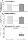
Stable isotope infusions were used to determine muscle protein net balance in a subgroup of 59 burned patients and 5 unburned young adults. Peripheral muscle protein synthesis was not altered at week 1 and week 3 when compared to unburned young adults (A). Protein breakdown however was increased 3–4 fold at 1 and 3 weeks postburn (B) leading to a negative protein net balance (C).
Figure 3
Figure 3
Severe burn causes marked changes in body composition during acute hospitalization (n=105). Severely burned children lost about 2% of their body weight, which is 5% lean body mass, 3% bone mineral content, 2% bone mineral density from admission to discharge. Total fat and percent fat increased from admission to discharge by 3% and 7%, respectively.
Figure 4
Figure 4
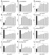
Serum complement C3 (A), α2-macroglobulin (B), haptoglobin (C), α1-acidglycoprotein (D), and CRP (E) were significantly increased postburn. Serum constitutive hepatic proteins pre-albumin (F), transferrin (G), retinol binding protein (H) markedly decreased immediately postburn and levels remained low up to 60 days postburn. Serum apolipoprotein A1 (I) significantly decreased postburn while apolipoprotein B (J) showed an increase. Serum free fatty acids (K) and triglycerides (L) significantly increased postburn. * Significant difference between burn vs. normal ranges, p<0.05.
Figure 5
Figure 5
Serum glucose increased during the acute phase postburn along with increased levels of endogenous insulin implying the presence of insulin resistance. * Significant difference between burned children vs. normal range, p<0.05.
Figure 6
Figure 6
Serum hormone levels. Serum IGF-I and IGFBP-3 (A–B) decreased markedly immediately after burn and remained significantly decreased throughout acute hospitalization. Serum GH (C) started to decrease 8–10 days postburn and showed a steady decline through acute hospitalization. Serum T4 (D) decreased immediately postburn but increased through acute hospitalization. Free thyroid index (FTI, E) showed a significant decrease for 14 days postburn and then returned to normal levels with no apparent difference. Serum cortisol (F) significantly increased immediately postburn and remained elevated for 3 weeks returning to normal levels. Urine cortisol (G) increased 5–7 folds during the acute stay but decreased over time. Serum osteocalcin (H) and iPTH (I) were drastically decreased (5–7 folds) immediately after burn and showed almost no increase over time. Serum β-estradiol (J) decreased immediately postburn but increased over time; testosterone (K) was normal during the early postburn phase, but showed marked decreases beginning 4 weeks postburn. Progesterone (L) was increased at various time points when compared with normal. * Significant difference between normal vs. burn, p<0.05.
Figure 7
Figure 7
All of the 17 serum cytokines measured were significantly altered (A) and these perturbations are possibly clinically relevantly. Dramatic changes were observed for serum G-CSF, IL-6, IL-8, MCP-1, and MIP-1β. * Significant difference between burn vs. normal ranges, p<0.05. Heat map (B) comparing normal (non-injured, non-burned children), and burned children controls at each time point <1 days postburn, (2–7 days postburn, 8–10 days postburn, 11–16 days postburn, 17–22 days postburn, 23–28 days postburn, 29–34 days postburn, and 35–60 days postburn. Values are log10 (average cytokine concentration, pg/ml); the color range for each cytokine is based on the detected values with blue indicating lower levels, yellow indicating highest levels, and black in the middle. Gray squares indicate that no expression was detected.
Figure 8
Figure 8
Cardiac output and predicted cardiac output was increased immediately postburn (up to 160% of predicted) and significantly decreased until discharge. Heart rate and predicted heart rate were also significantly increased postburn and remained elevated at discharge. Cardiac index was increased immediately postburn and significantly decreased from admission to discharge. * Significant difference between admission and discharge, p<0.05.
Figure 9
Figure 9
Immediately after burn, liver size doubled and at week 2, postburn was 220% of predicted liver size. Liver size remained elevated when patients were discharged from the ICU. * Significant difference between burn vs. normal range, p<0.05.

Similar articles

Cited by

References

    1. Przkora R, Barrow RE, Jeschke MG, et al. Body composition changes with time in pediatric burn patients. J Trauma. 2006;60:968–971. - PubMed
    1. Przkora R, Jeschke MG, Barrow RE, et al. Metabolic and hormonal changes of severely burned children receiving long-term oxandrolone treatment. Ann Surg. 2005;242:384–391. - PMC - PubMed
    1. Finnerty CC, Herndon DN, Przkora R, et al. Cytokine expression profile over time in severely burned pediatric patients. Shock. 2006;26:13–19. - PubMed
    1. Herndon DN, Tompkins RG. Support of the metabolic response to burn injury. Lancet. 2004;363:1895–1902. - PubMed
    1. Wilmore DW. Hormonal responses and their effect on metabolism. Surg Clin North Am. 1976;56:999–1018. - PubMed

Publication types

Associated data