. 2008 Sep 15;86(5):702-9.
doi: 10.1097/TP.0b013e31818410a3.

Expression of tissue factor and initiation of clotting by human platelets and monocytes after incubation with porcine endothelial cells

Affiliations

Expression of tissue factor and initiation of clotting by human platelets and monocytes after incubation with porcine endothelial cells

Chih Che Lin et al. Transplantation. .

Abstract

Objectives: Intravascular thrombosis remains a major barrier to successful pig-to-primate xenotransplantation. However, the precise factors initiating thrombosis are unknown. In this study, we investigated the contribution of recipient platelets and monocytes.

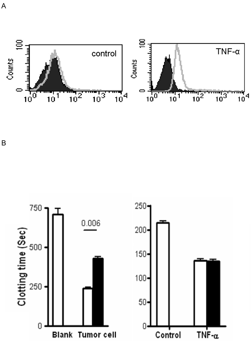
Methods: Primary pig aortic endothelial cells (PAECs) were incubated with combinations of fresh or heat-inactivated human plasma, platelets, or monocytes, after which they were separated and analyzed individually by flow cytometry for tissue factor (TF) expression and for their ability to clot recalcified normal or factor-VII-deficient plasma.

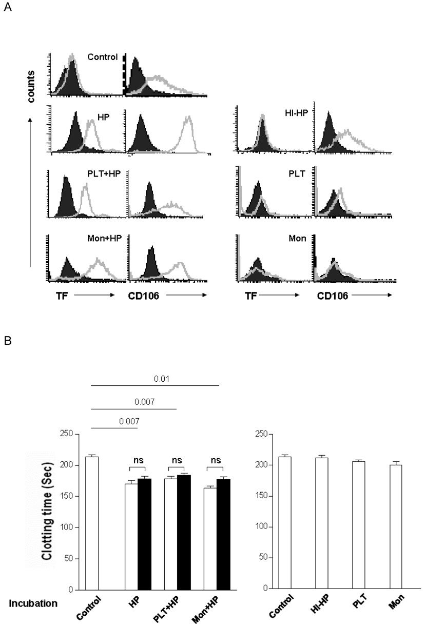
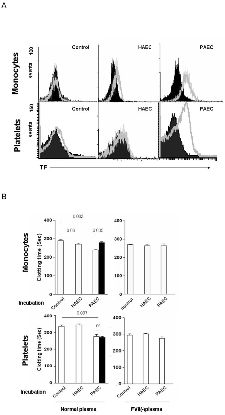
Results: Procoagulant porcine TF was induced in PAECs only by fresh human plasma, and not by heat-inactivated plasma, platelets, or monocytes. In contrast, procoagulant human TF was induced on platelets and monocytes after incubation with PAEC, irrespective of whether the plasma was present or not. In addition, human platelets caused the shedding of procoagulant TF-expressing aggregates from PAEC.

Conclusions: This work defines a cell-based in vitro assay system to address complex interactions among PAECs, human platelets, and monocytes. The induction of procoagulant TF on PAECs by fresh human plasma was most likely dependent on xenoreactive natural antibody and complement present in fresh human plasma. In contrast, the shedding of procoagulant platelet-PAEC aggregates, induced by human platelets, and the induction of procoagulant TF on human platelets and monocytes by PAEC, occurred independently of these factors. These results suggest that different mechanisms may contribute to the initiation of thrombosis after xenotransplantation, some of which may not be influenced by the further manipulation of the immune response against pig xenografts.


Figures

Figure 1
Figure 1. Characterisation of the sheep anti-human TF antibody
(A) PAEC stimulated by TNF-α (10ng/ml) for 8h to induce porcine TF activity measured by flow cytometery using the sheep anti-human TF antibody (open file) or an isotype control (closed file). (B) Clotting times of recalcified normal human plasma. Left panel; results without exposure to cells (control, blank) or after exposure to a human TF-positive tumor cell line. Right panel; results following no activation (control) or TNFα activation of PAEC. (Open bars - no additional sheep anti-human TF antibody. Closed bars - sheep anti-human TF antibody incubated with cells for 30min prior to the clotting assay.) (C) Recalcified coagulation assay using factor VII-deficient plasma.
Figure 2
Figure 2. PAEC express TF as determined by flow cytometry and demonstrate porcine TF activity in the recalcified clotting assay
PAEC were co-incubated with various stimuli for 8h, and studied by flow cytometery and the recalcified coagulation assay performed using both normal and factor VII-deficient plasma. Addition of sheep anti-human TF antibody was performed separately to determine the origin of the TF. (A) Results of flow cytometry of TF and CD106 expression (open files) on PAEC (closed files: isotype control). (B) Recalcified coagulation assay using normal human plasma with (open bars) or without (closed bars) incubation with sheep anti-human TF antibody after PAEC were pre-incubated in the presence (left) or absence (right) of fresh human plasma. (C) Recalcified coagulation assay using factor VII-deficient human plasma after PAEC were pre-incubated in the presence (left) or absence (right) of fresh human plasma. (ns = no significant difference, HP = human plasma, HI-HP = heated-inactivated human plasma, Mon = monocytes, PLT = platelets).
Figure 3
Figure 3. In the presence of human monocytes and platelets express human TF (after co-incubation with PAEC) as determined by flow cytometry, and demonstrate human TF activity in the recalcified clotting assay
Human monocytes and platelets were harvested from supernatants after co-incubation with HAEC or PAEC in the presence of 5% human plasma for 8h. (A) Results of flow cytometry of TF expression (open) on monocytes and platelets (closed files: isotype control). (B) Clotting times of recalcified normal (left) and factor VII-deficient (FVII(-)) (right) human plasma in the presence of resting (control) or pre-incubated (with HAEC+HP or PAEC+HP) human monocytes or platelets. Coagulation assays were also performed following prior incubation of the monocytes or platelets with sheep anti-human TF antibody (closed bars).
Figure 4
Figure 4. In the absence of human monocytes express human TF activity (after coincubation with PAEC), but only platelets confer porcine TF activity
Human monocytes and platelets were harvested from supernatants after co-incubation with PAEC in the absence of 5% human plasma for 8h. (A) Results of flow cytometry of TF expression on monocytes and platelets (open files) (closed files: isotype control). (B) Clotting times of recalcified normal (left) and factor VII-deficient (FVII(-)) (right) human plasma in the presence of resting (control) or pre-incubated (with HAEC or PAEC) human monocytes or platelets. Coagulation assays were also performed following prior incubation of the monocytes or platelets with the sheep anti-human TF antibody (closed bars). (ns = no significant difference).
Figure 5
Figure 5. Human platelets induce PAEC apoptosis
(A) PAEC were isolated after co-incubation with various stimuli for 8h. Apoptosis was measured by flow cytometry, double-staining with annexin V-FTIC and propidium iodide-PE defined apoptotic cells. The percentage of cells double-positive for TF and annexin V or single-positive for annexin V are indicated. (B) Immunocytochemical analysis of platelets following co-incubation with or without PAEC for 8h. Cells were double-stained with the sheep anti-human-TF antibody (FITC, green) and anti-porcine CD106 antibody (TRITC, red), then examined by fluorescence microscopy. TF-positive and CD106-negative cells are indicated by arrows. The larger, irregular particles double-positive for both markers are indicated by arrowheads; these were assumed to be fragments of apoptotic PAEC that expressed porcine TF (platelet-PAEC aggregates).

References

    1. Cooper DK, Gollackne B, Sachs DH. Will the pig solve the transplantation backlog? Annu Rev Med. 2002;53:133. - PubMed
    1. Cooper DK, Dorling A, Pierson RN, 3rd, et al. Alpha1,3-galactosyltransferase gene-knockout pigs for xenotransplantation: where do we go from here? Transplantation. 2007;84(1):1. - PubMed
    1. Phelps CJ, Koike C, Vaught TD, et al. Production of alpha 1,3-galactosyltransferase-deficient pigs. Science. 2003;299(5605):411. - PMC - PubMed
    1. Kuwaki K, Tseng YL, Dor FJ, et al. Heart transplantation in baboons using alpha1,3-galactosyltransferase gene-knockout pigs as donors: initial experience. Nat Med. 2005;11(1):29. - PubMed
    1. Tseng YL, Kuwaki K, Dor FJ, et al. alpha1,3-Galactosyltransferase gene-knockout pig heart transplantation in baboons with survival approaching 6 months. Transplantation. 2005;80(10):1493. - PubMed

Publication types