. 2009 Aug;66(8):437-47.
doi: 10.1002/cm.20316.

The ability to survive mitosis in the presence of microtubule poisons differs significantly between human nontransformed (RPE-1) and cancer (U2OS, HeLa) cells

Affiliations

The ability to survive mitosis in the presence of microtubule poisons differs significantly between human nontransformed (RPE-1) and cancer (U2OS, HeLa) cells

Daniela A Brito et al. Cell Motil Cytoskeleton. 2009 Aug.

Abstract

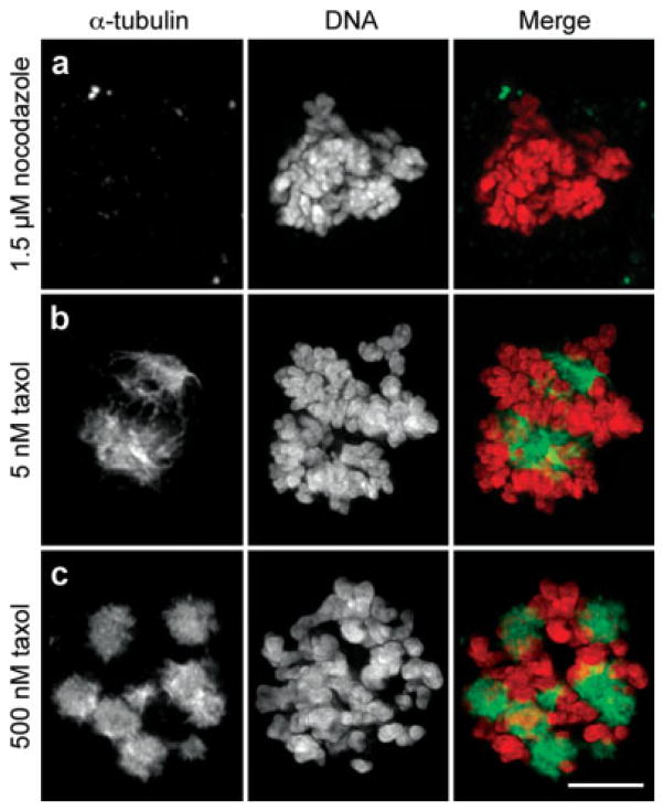
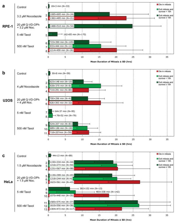
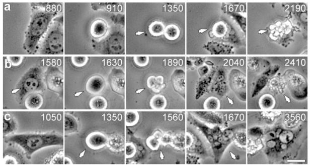
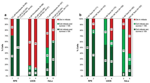
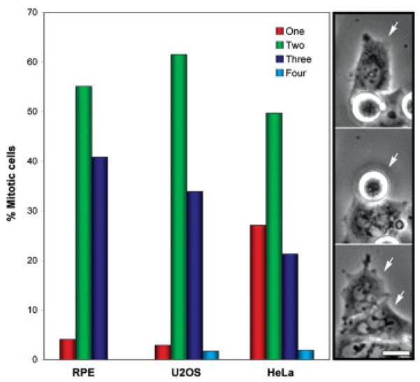
We used live cell imaging to compare the fate of human nontransformed (RPE-1) and cancer (HeLa, U2OS) cells as they entered mitosis in nocodazole or taxol. In the same field, and in either drug, a cell in all lines could die in mitosis, exit mitosis and die within 10 h, or exit mitosis and survive > or =10 h. Relative to RPE-1 cells, significantly fewer HeLa or U2OS cells survived mitosis or remained viable after mitosis: in nocodazole concentrations that inhibit spindle microtubule assembly, or in 500 nM taxol, 30% and 27% of RPE-1 cells, respectively, died in or within 10 h of exiting mitosis while 90% and 49% of U2OS and 78% and 81% of HeLa died. This was even true for clinically relevant taxol concentrations (5 nM) which killed 93% and 46%, respectively, of HeLa and U2OS cells in mitosis or within 10 h of escaping mitosis, compared to 1% of RPE-1 cells. Together these data imply that studies using HeLa or U2OS cells, harvested after a prolonged block in mitosis with nocodazole or taxol, are significantly contaminated with dead or dying cells. We also found that the relationship between the duration of mitosis and survival is drug and cell type specific and that lethality is related to the cell type and drug used to prevent satisfaction of the kinetochore attachment checkpoint. Finally, work with a pan-caspase inhibitor suggests that the primary apoptotic pathway triggered by nocodazole during mitosis in RPE-1 cells is not active in U2OS cells. Cell Motil. Cytoskeleton 2008. (c) 2008 Wiley-Liss, Inc.


Figures

Fig. 1
Fig. 1
Nocodazole inhibits spindle microtubule formation whereas taxol promotes microtubule stability. HeLa cells were incubated overnight with 1.5 μM nocodazole (a), 5 nM (b) taxol, or 500 nM (c) taxol prior to fixation and immunostaining for α-tubulin. First column, α-tubulin; second column, DNA; third column, merged images. HeLa that enter mitosis in the presence of 1.5 μM nocodazole fail to form spindle microtubules. The same is true for RPE-1 and U2OS cells treated, respectively, with 3.2 and 4.0 μM nocodazole (not shown). When exposed continuously to 5 nM taxol HeLa cells form abnormal but functional spindles that ultimately satisfy the kinetochore attachment checkpoint (b) while in 500 nM taxol multiple stable radial microtubule arrays (asters) are formed that the chromosomes attach to (c). The same is true for RPE-1 and U2OS (not shown). Bar = 5 μm.
Fig. 2
Fig. 2
In the presence of nocodazole or taxol, U2OS cells spend significantly less time in mitosis than RPE-1 or HeLa cells. Phase-contrast recordings of RPE-1 (a), U2OS (b) and HeLa (c) cultures continuously incubated with nocodazole or taxol in the presence or absence of a pancaspase inhibitor (Q-VD-OPh) were scored for cells that died in mitosis (red), died within 10 h of escaping mitosis (light green), or survived ≥10 h after escaping mitosis (dark green). Percentages were calculated from data pooled from ≥ 2 separate long-term recordings obtained on different days. Q-VD-OPh data are from single experiments. The white box in each bar defines the mean duration of mitosis in minutes ± the standard deviation, while N defines the total number of cells obtained in each category (see also Materials and Methods). See text for details.
Fig. 3
Fig. 3
Human cells in the same population that enter mitosis in the presence of microtubule poisons exhibit one of three fates. Selected frames from the same phase-contrast video recording of HeLa cells entering mitosis in 5 nM taxol. (a) This cell enters mitosis at 910 min, and then dies in mitosis at the 2190 min time point (arrow). (b) This cell enters mitosis at 1630 min, escapes mitosis (blebbing in 1890 min) to form a 4N cell (2040 min) that subsequently dies (2410 min) <10 h after exiting mitosis. (c) This cell enters mitosis at 1350 min (arrow), exits at 1560 min (arrow), and survives ≥10 h (until filming was terminated). Time, relative to addition of taxol, is shown in minutes. Bar = 10 μm.
Fig. 4
Fig. 4
Nontransformed RPE-1 cells show higher survival rates when dividing in the presence of nocodazole or taxol than U2OS or HeLa cells. Phase-contrast recordings of RPE-1, U2OS and HeLa cultures incubated with nocodazole (a) or taxol (b) were scored (cf. Fig. 2) for cells that died in mitosis (red), died within 10 h of escaping mitosis (light green), or survived ≥10 h after escaping mitosis (dark green). Numbers in the white box within each subset of an experimental treatment note the percentage of cells in that subset. (a) RPE-1, U2OS and HeLa cells treated with nocodazole alone (lanes 1, 3, and 5) or nocodazole and Q-VD-OPh (a caspase inhibitor; lanes 2, 4, and 6). Concentrations and total cell number are indicated above each column. Note that relative to RPE-1 cells, nocodazole concentrations that inhibit spindle MT assembly kill substantially more U2OS and HeLa cells during or shortly after mitosis. (b) RPE-1, U2OS and HeLa cultures treated with either 5 nM (lanes 1, 3, and 5) or 500 nM (lanes 2, 4, and 6) taxol. As for nocodazole, compared to RPE-1 cells U2OS and HeLa have a more difficult time surviving mitosis in the presence of taxol. Note also that taxol kills substantially fewer U2OS than HeLa cells during or after mitosis, and that only 7% of HeLa cells dividing in 5 nM taxol are alive 10 h later.
Fig. 5
Fig. 5
Low magnification time-lapse recordings of RPE-1, U2OS and HeLa cells dividing in the presence of 5 nM taxol were analyzed for cells that exited mitosis without dividing (red bars), or that exited mitosis and divided into two (green bars), three (blue bars) or four daughters (light blue bars). Images on the right depict selected frames of a HeLa cell that divided in the presence of 5 nM taxol into two daughters (white arrows in bottom frame) both of which were micro-nucleated. Bar = 10 μm.

References

    1. Allan LA, Clarke PR. Phosphorylation of caspase-9 by CDK1/cyclin B1 protects mitotic cells against apoptosis. Mol Cell. 2007;26:301–310. - PubMed
    1. Andreassen PR, Margolis RL. Microtubule dependency of p34cdc2 inactivation and mitotic exit in mammalian cells. J Cell Biol. 1994;127:789–802. - PMC - PubMed
    1. Brito DA, Rieder CL. Mitotic checkpoint slippage in vertebrates occurs via cyclin B destruction in the presence of an active checkpoint. Curr Biol. 2006;16:1194–1200. - PMC - PubMed
    1. Brito DA, Yang Z, Rieder CL. Microtubules do not promote mitotic slippage when the spindle assembly checkpoint cannot be satisfied. J Cell Biol. 2008;182:623–629. - PMC - PubMed
    1. Cahill DP, Lengauer C, Yu J, Riggins GJ, Wilson JK, Markowitz SD, Kinzler KW, Vogelstein B. Mutations of mitotic checkpoint genes in human cancers. Nature. 1998;392:300–303. - PubMed

Publication types

LinkOut - more resources