. 2008 Nov;15(6):707-17.
doi: 10.1007/s11373-008-9278-3. Epub 2008 Sep 16.

Interactions between M protein and other structural proteins of severe, acute respiratory syndrome-associated coronavirus

Affiliations

Interactions between M protein and other structural proteins of severe, acute respiratory syndrome-associated coronavirus

Yi-Ching Hsieh et al. J Biomed Sci. 2008 Nov.

Abstract

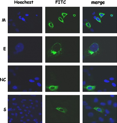
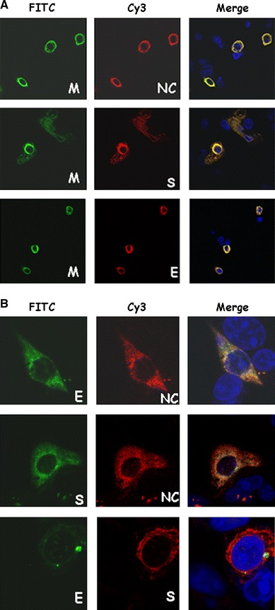
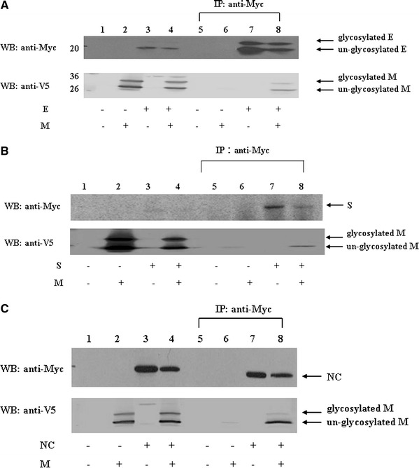
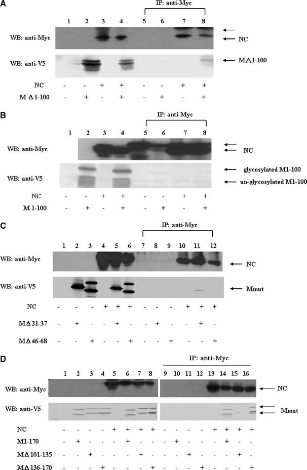
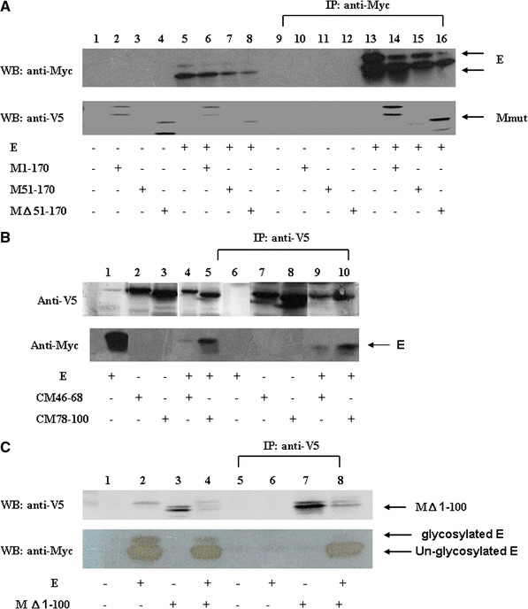
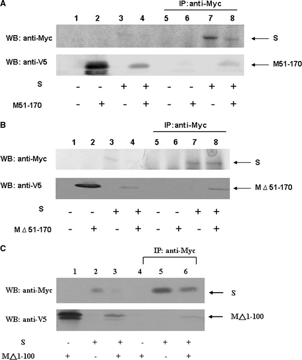
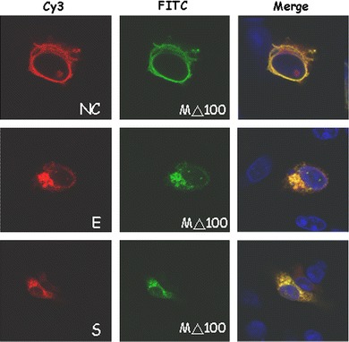
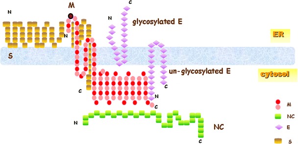
Severe acute respiratory syndrome-associated coronavirus (SARS-CoV) structural proteins (S, E, M, and NC) localize in different subcellular positions when expressed individually. However, SARS-CoV M protein is co-localized almost entirely with S, E, or NC protein when co-expressed in the cells. On the other hand, only partial co-localization was observed when S and E, S and NC, or E and NC were co-expressed in the cells. Interactions between SARS-CoV M and other structural proteins but not interactions between S and E, S and NC, or E and NC were further demonstrated by co-immunoprecipitation assay. These results indicate that SARS-CoV M protein, similar to the M proteins of other coronaviruses, plays a pivotal role in virus assembly. The cytoplasmic C-terminus domain of SARS-CoV M protein was responsible for binding to NC protein. Multiple regions of M protein interacted with E and S proteins. A model for the interactions between SARS-CoV M protein and other structural proteins is proposed. This study helps us better understand protein-protein interactions during viral assembly of SARS-CoV.


Figures

Fig. 1
Fig. 1
Differential subcellular localizations of SARS-CoV structural proteins S, E, M and NC) when they were expressed individually. Cells were transfected with the plasmid expressing M-V5, the plasmid expressing myc-E protein, the plasmid expressing NC protein, or the plasmid expressing S protein. After transfection, cells were fixed and stained with mouse anti-V5, mouse anti-myc, rabbit anti-NC, rabbit anti-S antibodies. Green color, M, NC, S, or E protein staining; blue color, DAPI staining
Fig. 2
Fig. 2
(a) Co-localization of SARS-CoV M protein with S, E, or NC protein when co-expressed in Vero E6 cells. Cells were co-transfected with plasmids expressing the M-V5 and NC (upper), S (middle), or myc-E (lower) proteins. After transfection, cells were fixed and stained with rabbit anti-NC and mouse anti-V5 antibodies (upper), or rabbit anti-S and mouse anti-V5 antibodies (middle), or mouse anti-myc and goat anti-mouse conjugated with Cy3 followed by anti-V5-FITC antibody (lower). Green color, M protein staining; red color, NC (or S, or E) protein staining; blue color, DAPI staining; yellow color, co-localization of M and other structural proteins. (b) Partial co-localization of E and NC, S and NC, or E and S proteins when they were co-expressed in Vero E6 cells. Cells were co-transfected with plasmids expressing myc-E and NC proteins (upper); S and myc-NC proteins (middle), and myc-E and S proteins (lower). After transfection, cells were fixed and stained with rabbit anti-NC (or anti-S) and mouse anti-myc antibodies
Fig. 3
Fig. 3
(a) SARS-CoV M interaction with E protein. Vero E6 cells were transfected with vector alone (lanes 1 and 5), plasmid encoding M-V5 (lanes 2 and 6), plasmid encoding myc-E (lanes 3 and 7), or co-transfected with both plasmids (lanes 4 and 8). Cell lysates were directly analyzed by Western blotting (lanes 1–4) or immunoprecipitated with the anti-myc antibody prior to Western blotting (lanes 5–8). (b) SARS-CoV M interaction with S proteins. Vero E6 cells were transfected with vector alone (lanes 1 and 5), plasmid encoding M-V5 (lanes 2 and 6), plasmid encoding myc-S (lanes 3 and 7), or co-transfected with both plasmids (lanes 4 and 8). Cell lysates were directly analyzed by Western blotting (lanes 1–4) or immunoprecipitated with the anti-myc antibody prior to Western blotting (lanes 5–8). (c) SARS-CoV M interaction with NC proteins. Vero E6 cells were transfected with vector alone (lanes 1 and 5), plasmid encoding M-V5 (lanes 2 and 6), plasmid encoding myc-NC (lanes 3 and 7), or co-transfected with both plasmids (lanes 4 and 8). Cell lysates were directly analyzed by Western blotting (lanes 1–4) or immunoprecipitated with the anti-myc antibody prior to Western blotting (lanes 5–8)
Fig. 4
Fig. 4
Immunoprecipitation and Western blotting analyses of SARS-CoV M protein cytoplasmic C-terminus domain interaction with NC protein. (a) Vero E6 cells were transfected with vector alone (lanes 1 and 5), plasmid encoding cytoplasmic C-terminus domain of M protein (i.e. M protein without its first 100 amino acids) with a V5 tag (lanes 2 and 6), plasmid encoding myc-NC (lanes 3 and 7), or co-transfected with both plasmids (lanes 4 and 8). Cell lysates were directly analyzed by Western blotting (lanes 1–4) or immunoprecipitated with the anti-myc antibody prior to Western blotting (lanes 5–8). The cytoplasmic C-terminus domain of M protein interacted with NC protein. The protein larger than NC protein marked by the thin arrow is the immunoglobulin heavy chain (lower panel). More than one band was detected when the plasmid encoding the cytoplasmic C-terminus domain of M protein was expressed, possibly due to sample preparation without boiling treatment. (b) Vero E6 cells were transfected with vector alone (lanes 1 and 5), plasmid encoding the first 100 amino acids of M protein with a V5 tag (lanes 2 and 6), plasmid encoding myc-NC (lanes 3 and 7), or co-transfected with both plasmids (lanes 4 and 8). Cell lysates were directly analyzed by Western blotting (lanes 1–4) or immunoprecipitated with the anti-myc antibody prior to Western blotting (lanes 5–8). M protein without its cytoplasmic domain did not interact with NC protein. The protein larger than NC protein marked by the thin arrow is the immunoglobulin heavy chain. (c) Vero E6 cells were transfected with vector alone, plasmid encoding M△21–37 plus a V5 tag (or the plasmid encoding M△46–68 plus a V5 tag), plasmid encoding myc-NC, or co-transfected with two plasmids (M△21–37 and myc-NC, M△46–68 and myc-NC). Cell lysates were directly analyzed by Western blotting (lanes 1–6) or immunoprecipitated with the anti-myc antibody prior to Western blotting (lanes 7–12). M protein without its first hydrophobic domain, but not M protein without its second hydrophobic domain interacted with NC protein. (d) Vero E6 cells were transfected with vector alone, plasmid encoding M1–170 with a V5 tag (or the plasmid encoding M△101–135 plus a V5 tag, or the plasmid encoding M△136–170 plus a V5 tag), plasmid encoding myc-NC, or co-transfected with two plasmids (M1–170 and myc-NC, M△101–135 and myc-NC, M△136–170 and myc-NC). Cell lysates were directly analyzed by Western blotting (lanes 1–8) or immunoprecipitated with the anti-myc antibody prior to Western blotting (lanes 9–18). All of the M cytoplasmic domain-deleted mutants interacted with NC protein
Fig. 5
Fig. 5
Immunoprecipitation and Western blotting analyses of SARS-CoV M protein fragment interactions with E protein. (a) Interactions between SARS-CoV E and different M fragments. Vero E6 cells were transfected with vector alone, with the plasmid encoding M1–170 with a V5 tag (or the plasmid encoding M51–170 plus a V5 tag, or the plasmid encoding M△51–170 plus a V5 tag), plasmid encoding myc-E, or co-transfected with two plasmids (M1–170 and myc-E, M51–170 and myc-E, M△51–170 and myc-E). Cell lysates were directly analyzed by Western blotting (lanes 1–8) or immunoprecipitated with the anti-myc antibody prior to Western blotting (lanes 9–18). (b) Vero E6 cells were transfected with the plasmid encoding myc-E protein, plasmid encoding the first 115 amino acids of HCV core protein and M protein amino acids 46–68 plus a V5 tag (the plasmid encoding the first 115 amino acids of HCV core protein and M protein amino acids 78–100 plus a V5 tag), or co-transfected with two plasmids. Cell lysates were directly analyzed by Western blotting (lanes 1–4) or immunoprecipitated with the anti-V5 antibody prior to Western blotting (lanes 5–8). Either the second or the third transmembrane domain of M protein was sufficient for interaction with E protein. (c) Interactions between SARS-CoV E and the cytoplasmic C-terminus domain of M protein. Vero E6 cells were transfected with vector alone, plasmid encoding myc-E protein, plasmid encoding M protein without the first 100 amino acids plus a V5 tag, or co-transfected with both plasmids. Cell lysates were directly analyzed by Western blotting (lanes 1–4) or immunoprecipitated with the anti-V5 antibody prior to Western blotting (lanes 5–8) (lower panel). More than one band was detected when the plasmid encoding the cytoplasmic C-terminus domain of M protein was expressed, possibly due to sample preparation without boiling treatment
Fig. 6
Fig. 6
M protein interaction with S protein. (a) Vero E6 cells were transfected with vector alone (lanes 1 and 5), plasmid encoding M51–170 with a V5 tag (lanes 2 and 6), plasmid encoding myc-S (lanes 3 and 7), or co-transfected with both plasmids (lanes 4 and 8). Cell lysates were directly analyzed by Western blotting (lanes 1–4) or immunoprecipitated with the anti-myc antibody prior to Western blotting (lanes 5–8). (b) Similar to (a), except the plasmid encoding M△51–170 plus a V5 tag was used to replace the plasmid encoding M51–170 with a V5 tag. (c) Interactions between SARS-CoV S and cytoplasmic C-terminus domain of M protein. Similar to (a), except the plasmid encoding M protein without the first 100 amino acids plus a V5 tag was used to replace the plasmid encoding M51–170 with a V5 tag (lower panel). More than one band was detected when the plasmid encoding cytoplasmic domain of M protein was expressed possibly due to the sample preparation without boiling treatment. The results show that multiple regions of M protein interacted with S protein
Fig. 7
Fig. 7
Co-localization of M△1–100 protein and NC (upper panel), E (middle panel), and S (lower panel) proteins. Similar to Fig. 2a, except the plasmid encoding M protein without the first 100 amino acids plus a V5 tag was used to replace the plasmid encoding M protein with a V5 tag
Fig. 8
Fig. 8
A proposed model for the interactions between the M protein and other structural proteins of SARS-CoV. M protein probably interacts with unglycosylated E protein through cytoplasmic C-terminus regions first. After glycosylation of E protein, interaction between M and E proteins are possibly through transmembrane regions, and the freed cytoplasmic region of M protein will then interact with NC protein. S protein could incorporate into virus-like particles formed by M and E proteins through interacting with multiple regions of M protein

Similar articles

Cited by

References

    1. Poon LL, Guan Y, Nicholls JM, Yuen KY, Peiris JS. The aetiology, origins, and diagnosis of severe acute respiratory syndrome. Lancet Infect Dis. 2004;4:663–671. doi: 10.1016/S1473-3099(04)01172-7. - DOI - PMC - PubMed
    1. Rota PA, Oberste MS, Monroe SS, Nix WA, Campagnoli R, Icenogle JP, Penaranda S, Bankamp B, Maher K, Chen MH, Tong S, Tamin A, Lowe L, Frace M, DeRisi JL, Chen Q, Wang D, Erdman DD, Peret TC, Burns C, Ksiazek TG, Rollin PE, Sanchez A, Liffick S, Holloway B, Limor J, McCaustland K, Olsen-Rasmussen M, Fouchier R, Gunther S, Osterhaus AD, Drosten C, Pallansch MA, Anderson LJ, Bellini WJ. Characterization of a novel coronavirus associated with severe acute respiratory syndrome. Science. 2003;300:1394–1399. doi: 10.1126/science.1085952. - DOI - PubMed
    1. Peiris JS, Lai ST, Poon LL, Guan Y, Yam LY, Lim W, Nicholls J, Yee WK, Yan WW, Cheung MT, Cheng VC, Chan KH, Tsang DN, Yung RW, Ng TK, Yuen KY. Coronavirus as a possible cause of severe acute respiratory syndrome. Lancet. 2003;361:1319–1325. - PMC - PubMed
    1. Kuiken T, Fouchier RA, Schutten M, Rimmelzwaan GF, van Amerongen G, van Riel D, Laman JD, de Jong T, van Doornum G, Lim W, Ling AE, Chan PK, Tam JS, Zambon MC, Gopal R, Drosten C, van der Werf S, Escriou N, Manuguerra JC, Stohr K, Peiris JS, Osterhaus AD. Newly discovered coronavirus as the primary cause of severe acute respiratory syndrome. Lancet. 2003;362:263–270. doi: 10.1016/S0140-6736(03)13967-0. - DOI - PMC - PubMed
    1. Gu J, Gong E, Zhang B, Zheng J, Gao Z, Zhong Y, Zou W, Zhan J, Wang S, Xie Z, Zhuang H, Wu B, Zhong H, Shao H, Fang W, Gao D, Pei F, Li X, He Z, Xu D, Shi X, Anderson VM, Leong AS. Multiple organ infection and the pathogenesis of SARS. J Exp Med. 2005;202:415–424. doi: 10.1084/jem.20050828. - DOI - PMC - PubMed

Publication types

MeSH terms

LinkOut - more resources