. 2009 May;127(1):123-33.
doi: 10.1111/j.1365-2567.2008.02929.x.

Regulation of macrophage nitric oxide production by the protein tyrosine phosphatase Src homology 2 domain phosphotyrosine phosphatase 1 (SHP-1)

Affiliations

Regulation of macrophage nitric oxide production by the protein tyrosine phosphatase Src homology 2 domain phosphotyrosine phosphatase 1 (SHP-1)

Julie Blanchette et al. Immunology. 2009 May.

Abstract

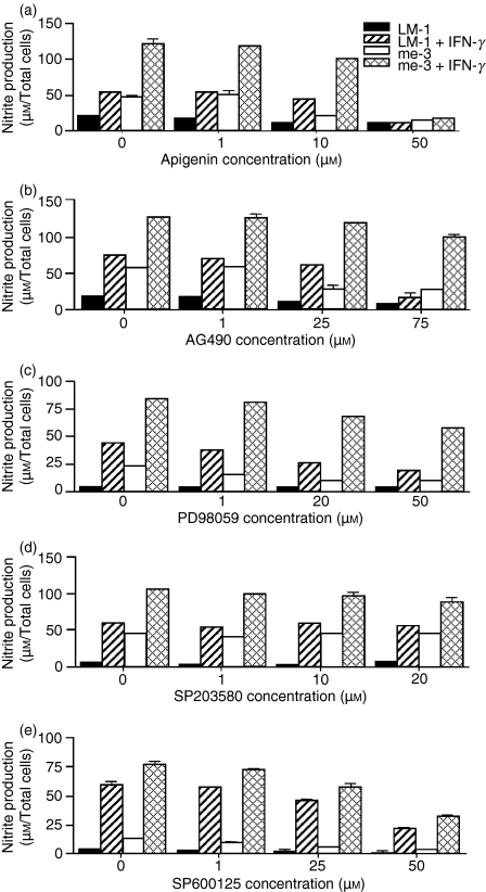
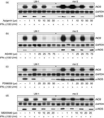
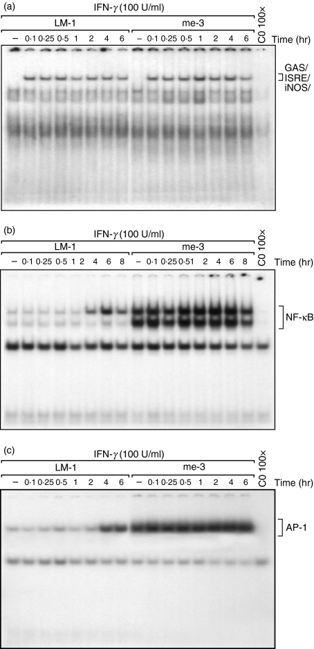
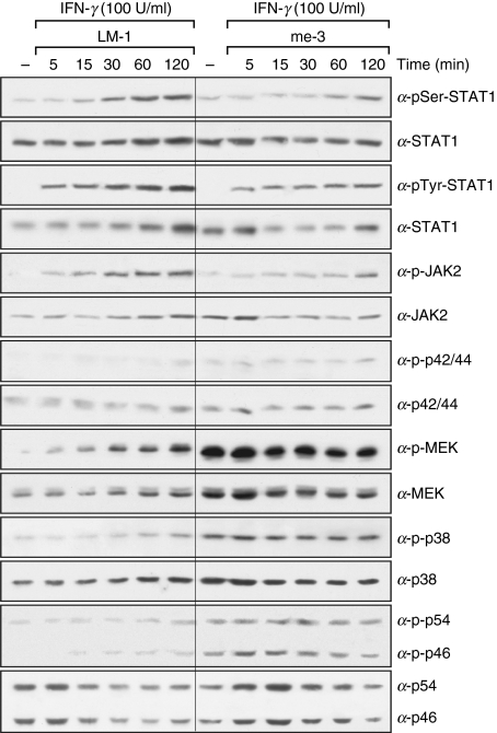
Nitric oxide (NO) is a potent molecule involved in the cytotoxic effects mediated by macrophages (MØ) against microorganisms. We previously reported that Src homology 2 domain phosphotyrosine phosphatase 1 (SHP-1)-deficient cells generate a greater amount of NO than wild-type cells in response to interferon-gamma (IFN-gamma). We also reported that the Leishmania-induced MØ SHP-1 activity is needed for the survival of the parasite within phagocytes through the attenuation of NO-dependent and NO-independent mechanisms. In the present study, we investigated the role of SHP-1 in regulating key signalling molecules important in MØ NO generation. Janus tyrosine kinase 2 (JAK2), mitogen-activated extracellular signal-regulated protein kinase kinase (MEK), extracellular signal-regulated kinases 1 and 2 (Erk1/Erk2) mitogen-activated protein kinases, p38 and stress-activated mitogen-activated protein kinases/c-Jun NH(2)-terminal kinase (SAPK/JNK) were examined in immortalized bone marrow-derived MØ (BMDM) from both SHP-1-deficient motheaten mice (me-3) and their respective littermates (LM-1). The results indicated that Erk1/Erk2 and SAPK/JNK are the main kinases regulated by SHP-1 because the absence of SHP-1 caused an increase in their phosphorylation. Moreover, only Apigenin, the specific inhibitor of Erk1/Erk2, was able to block IFN-gamma-induced inducible nitric oxide synthase (iNOS) transcription and translation in me-3 cells. Transcription factor analyses revealed that in the absence of SHP-1, activator protein-1 (AP-1) was activated. The activation of AP-1, and not nuclear factor-kappaB (NF-kappaB) or signal transducer and activator of transcription-1 alpha (STAT-1 alpha), may explain the enhanced NO generation in SHP-1-deficient cells. These observations emphasize the involvement of the MAPKs Erk1/Erk2 and SAPK/JNK in NO generation via AP-1 activation. Collectively, our findings suggest that SHP-1 plays a pivotal role in the negative regulation of signalling events leading to iNOS expression and NO generation. Furthermore, our observations underline the importance of SHP-1-mediated negative regulation in maintaining NO homeostasis and thus preventing the abnormal generation of NO that can be detrimental to the host.


Figures

Figure 1
Figure 1
Effect of the specific inhibitors of Janus tyrosine kinase 2 (JAK2), extracellular signal-regulated kinases 1 and 2 (Erk1/Erk2), mitogen-activated extracellular signal-regulated protein kinase kinase (MEK), p38 and stress-activated mitogen-activated protein kinases/c-Jun NH2-terminal kinase (SAPK/JNK) on nitric oxide (NO) generation in Src homology 2 domain phosphotyrosine phosphatase 1 (SHP-1)-deficient cells (me-3) and their wild-type counterparts (LM-1). Macrophages (MØ) were incubated with increasing doses of the Erk1/Erk2 inhibitor (Apigenin; 1–50 μm) (a), the JAK2 inhibitor (AG490; 1–75 μm) (b), the MEK inhibitor (PD98059, 1–40 μm) (c), the p38 inhibitor (SB203580; 1–20 μm) (d) and the SAPK/JNK inhibitor (SP600125), 1–50 μm) (e) for 1 hr prior to stimulation with interferon-γ (IFN-γ) (100 U/ml, 24 hr). Generation of NO was measured as described in the Experimental procedures. The results are representative of three independent experiments.
Figure 2
Figure 2
Effect of the kinase inhibitors on the expression of inducible nitric oxide synthase (iNOS) in Src homology 2 domain phosphotyrosine phosphatase 1 (SHP-1)-deficient cells (me-3) in response to interferon-γ (IFN-γ). Cells were incubated with increasing doses of the extracellular signal-regulated kinases 1 and 2 (Erk1/Erk2) inhibitor Apigenin (1–50 μm) (a), Janus tyrosine kinase 2 (JAK2) inhibitor AG490 (1–75 μm) (b), mitogen-activated extracellular signal-regulated protein kinase kinase (MEK) inhibitor PD98059 (1–40 μm) (c), or the p38 inhibitor SB203580 (1–20 μm) (d), for 1 hr prior to stimulation with IFN-γ. Expression of the iNOS gene (8 hr poststimulation) and iNOS protein (24 hr poststimulation) were monitored by Northern and western blot analyses, respectively, as described in the Experimental procedures. The results are representative of three independent experiments. GAPDH, glyceraldehyde 3-phosphate dehydrogenase.
Figure 3
Figure 3
Modulation of interferon-γ (IFN-γ)-mediated nuclear translocation of signal transducer and activator of transcription 1α (STAT-1α), nuclear factor-κB (NF-κB) and activator protein-1 (AP-1) transcription factors in Src homology 2 domain phosphotyrosine phosphatase 1 (SHP-1)-deficient cells (me-3) and their wild-type counterparts (LM-1). Cells were stimulated with IFN-γ for different periods of time (0–8 hr). Activation of STAT-1α was monitored using an oligonucleotide for the specific binding site on inducible nitric oxide synthase (iNOS) (GAS/ISRE/iNOS) (a). Nuclear translocation was also monitored using specific oligonucleotides for NF-κB (b) and AP-1 (c). The last lane in each panel represents nuclear extracts of me-3 cells stimulated with IFN-γ for 1 hr and subjected to competition with the respective cold oligonucleotide to confirm signal specificity. The results are representative of three independent experiments.
Figure 4
Figure 4
Role of nuclear factor-κB (NF-κB) in nitric oxide (NO) generation and inducible nitric oxide synthase (iNOS) expression in interferon-γ (IFN-γ)-stimulated Src homology 2 domain phosphotyrosine phosphatase 1 (SHP-1)-deficient cells (me-3) and their wild-type counterparts (LM-1). (a) Both cell lines were treated with two different NF-κB inhibitors, namely sodium salicylate (NaS, 5 mm) and BAY 11-7082 (5 μm) for 1 hr prior to stimulation with IFN-γ (100 U/ml). After 24 hr of incubation in the presence of the inhibitor, supernatants were collected and the Greiss reaction was performed to evaluate nitrite generation. The results with both inhibitors represent three independent experiments (mean ± standard error of the mean, n = 3). (b) Expression of the iNOS gene was monitored in cells treated for 1 hr with increasing doses of both inhibitors (1–5 mm NaS and 1–5 μm BAY 11-7082) prior to stimulation with IFN-γ (100 U/ml, 8 hr). Inhibitors were retained throughout the IFN-γ stimulation period. The results are representative of three independent experiments. GAPDH, glyceraldehyde 3-phosphate dehydrogenase.
Figure 5
Figure 5
Time-dependent activation of signal transducer and activator of transcription 1α (STAT-1α), Janus tyrosine kinase 2 (JAK2), extracellular signal-regulated kinases 1 and 2 (Erk1/Erk2), mitogen-activated extracellular signal-regulated protein kinase kinase (MEK), p38 and stress-activated mitogen-activated protein kinases/c-Jun NH2-terminal kinase (SAPK/JNK) in Src homology 2 domain phosphotyrosine phosphatase 1 (SHP-1)-deficient cells (me-3) and their wild-type counterparts (LM-1) stimulated with interferon-γ (IFN-γ). Cells were treated with IFN-γ (100 U/ml) for various periods of time (5–120 min). Cell lysates were subjected to western blot analyses using antibodies specific for the phosphorylated forms of STAT-1α seryl 727 and tyrosyl 701 residues (α-pSer-STAT1 and α-pTyr-STAT1, respectively), JAK2 (α-p-JAK2), Erk1/Erk2 MAPKs (α-p-p44 and α-p-p42), MEK (α-p-MEK), p38 (α-p-p38) and SAPK/JNK MAPK (α-p-p54 and α-p-p46). Loading of equal amounts of protein was monitored by stripping the membranes and reblotting with antibodies specific for the non-phosphorylated form of the various kinases analyzed. The results are representative of three independent experiments.
Figure 6
Figure 6
Effect of the extracellular signal-regulated kinases 1 and 2 (Erk1/Erk2) and p38 inhibitors on activator protein-1 (AP-1) nuclear translocation in interferon-γ (IFN-γ)-stimulated Src homology 2 domain phosphotyrosine phosphatase 1 (SHP-1)-deficient cells (me-3) and their wild-type counterparts (LM-1). The effect of the Erk1/Erk2 inhibitor Apigenin (50 μm) and p38 inhibitor SB203580 (20 μm) (both added for 1 hr prior to IFN-γ and retained throughout the IFN-γ stimulation period) on AP-1 nuclear translocation was evaluated in cells stimulated for 4 hr with IFN-γ or left unstimulated. AP-1 activation was monitored as described in the Experimental procedures. The results are representative of three independent experiments. CO, cold oligo.

References

    1. Green SJ, Nacy CA, Meltzer MS. Cytokine-induced synthesis of nitrogen-oxides in macrophages – a protective host response to Leishmania and other intracellular pathogens. J Leukoc Biol. 1991;50:93–103. - PubMed
    1. Liew FY. The role of nitric-oxide in parasitic diseases. Ann Trop Med Parasitol. 1993;87:637–42. - PubMed
    1. Lowenstein CJ, Dinerman JL, Snyder SH. Nitric-oxide – a physiological messenger. Ann Intern Med. 1994;120:227–37. - PubMed
    1. Croen KD. Evidence for an antiviral effect of nitric-oxide – inhibition of Herpes-Simplex Virus type-1 replication. J Clin Invest. 1993;91:2446–52. - PMC - PubMed
    1. Karupiah G, Xie QW, Buller RML, Nathan C, Duarte C, Macmicking JD. Inhibition of viral replication by interferon-gamma-induced nitric-oxide synthase. Science. 1993;261:1445–8. - PubMed

Publication types

MeSH terms