Comparative Study
. 2008 Sep 15:8:83.
doi: 10.1186/1471-213X-8-83.

The Mouse Limb Anatomy Atlas: an interactive 3D tool for studying embryonic limb patterning

Affiliations
Comparative Study

The Mouse Limb Anatomy Atlas: an interactive 3D tool for studying embryonic limb patterning

April Delaurier et al. BMC Dev Biol. .

Abstract

Background: The developing mouse limb is widely used as a model system for studying tissue patterning. Despite this, few references are available that can be used for the correct identification of developing limb structures, such as muscles and tendons. Existing textual references consist of two-dimensional (2D) illustrations of the adult rat or mouse limb that can be difficult to apply when attempting to describe the complex three-dimensional (3D) relationship between tissues.

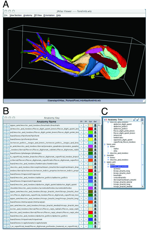
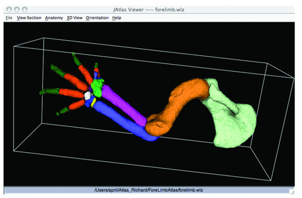
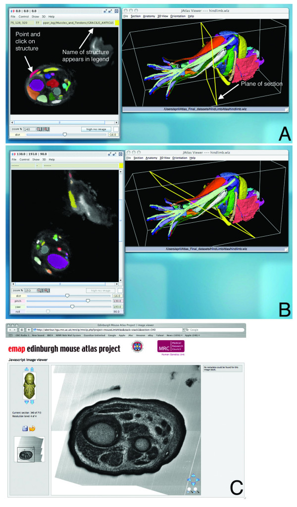
Results: To improve the resources available in the mouse model, we have generated a free, web-based, interactive reference of limb muscle, tendon, and skeletal structures at embryonic day (E) 14.5 http://www.nimr.mrc.ac.uk/3dlimb/. The Atlas was generated using mouse forelimb and hindlimb specimens stained using immunohistochemistry to detect muscle and tendon. Limbs were scanned using Optical Projection Tomography (OPT), reconstructed to make 3D models and annotated using computer-assisted segmentation tools in Amira 3D Visualisation software. The annotated dataset is visualised using Java, JAtlasView software. Users click on the names of structures and view their shape, position and relationship with other structures within the 3D model and also in 2D virtual sections.

Conclusion: The Mouse Limb Anatomy Atlas provides a novel and valuable tool for researchers studying limb development and can be applied to a range of research areas, including the identification of abnormal limb patterning in transgenic lines and studies of models of congenital limb abnormalities. By using the Atlas for "virtual" dissection, this resource offers an alternative to animal dissection. The techniques we have developed and employed are also applicable to many other model systems and anatomical structures.


Figures

Figure 1
Figure 1
JAtlasView. A: JAtlasView showing an interactive 3D model of the muscles, tendons and bones of the embryonic mouse forelimb at E14.5. B: Anatomy key. Shows the name of each structure in the 3D model, its ontogeny and colour code. Structures can be switched off/on in the viewer window by clicking on the '+/-' icon in the Anatomy Key. C: Anatomy Tree. Shows the ontogeny of structures. Names can be clicked so structures appear in the 3D model.
Figure 2
Figure 2
JAtlasView showing an interactive 3D model of the skeletal structures of the embryonic mouse forelimb at E14.5 after switching off muscles and tendons in the Anatomy Key.
Figure 3
Figure 3
JAtlasView showing 2D section views through the hindlimb. A: XY section view. Using the point-and-click function of the 2D viewer, structures can be clicked on in the section and the name appears at the top of the 2D window. The plane of section is visible in the 3D view. B: A user-defined section. The plane of section is changed by adjusting 'yaw' and 'pitch'. C: High-resolution section viewer. By clicking on "high-res image" in the 2D viewer, the Java Section Viewer is launched. Users can move arrows to pan through the stack, magnify and translate sections. This section is equivalent to the section shown in 3A.

References

    1. Kerwin J, Scott M, Sharpe J, Puelles L, Robson SC, Martinez-de-la-Torre M, Ferran JL, Feng G, Baldock R, Strachan T, et al. 3 dimensional modelling of early human brain development using optical projection tomography. BMC Neurosci. 2004;5:27. doi: 10.1186/1471-2202-5-27. - DOI - PMC - PubMed
    1. Sharpe J, Ahlgren U, Perry P, Hill B, Ross A, Hecksher-Sorensen J, Baldock R, Davidson D. Optical projection tomography as a tool for 3D microscopy and gene expression studies. Science. 2002;296:541–5. doi: 10.1126/science.1068206. - DOI - PubMed
    1. Weninger WJ, Mohun T. Phenotyping transgenic embryos: a rapid 3-D screening method based on episcopic fluorescence image capturing. Nat Genet. 2002;30:59–65. doi: 10.1038/ng785. - DOI - PubMed
    1. Rosenthal J, Mangal V, Walker D, Bennett M, Mohun TJ, Lo CW. Rapid high resolution three dimensional reconstruction of embryos with episcopic fluorescence image capture. Birth Defects Res C Embryo Today. 2004;72:213–23. doi: 10.1002/bdrc.20023. - DOI - PubMed
    1. Goncalves LF, Espinoza J, Romero R, Kusanovic JP, Swope B, Nien JK, Erez O, Soto E, Treadwell MC. Four-dimensional ultrasonography of the fetal heart using a novel Tomographic Ultrasound Imaging display. J Perinat Med. 2006;34:39–55. doi: 10.1515/JPM.2006.006. - DOI - PMC - PubMed

Publication types

LinkOut - more resources