. 2008 Sep 15:5:38.
doi: 10.1186/1742-2094-5-38.

Sepsis causes neuroinflammation and concomitant decrease of cerebral metabolism

Affiliations

Sepsis causes neuroinflammation and concomitant decrease of cerebral metabolism

Alexander Semmler et al. J Neuroinflammation. .

Abstract

Background: Septic encephalopathy is a severe brain dysfunction caused by systemic inflammation in the absence of direct brain infection. Changes in cerebral blood flow, release of inflammatory molecules and metabolic alterations contribute to neuronal dysfunction and cell death.

Methods: To investigate the relation of electrophysiological, metabolic and morphological changes caused by SE, we simultaneously assessed systemic circulation, regional cerebral blood flow and cortical electroencephalography in rats exposed to bacterial lipopolysaccharide. Additionally, cerebral glucose uptake, astro- and microglial activation as well as changes of inflammatory gene transcription were examined by small animal PET using [18F]FDG, immunohistochemistry, and real time PCR.

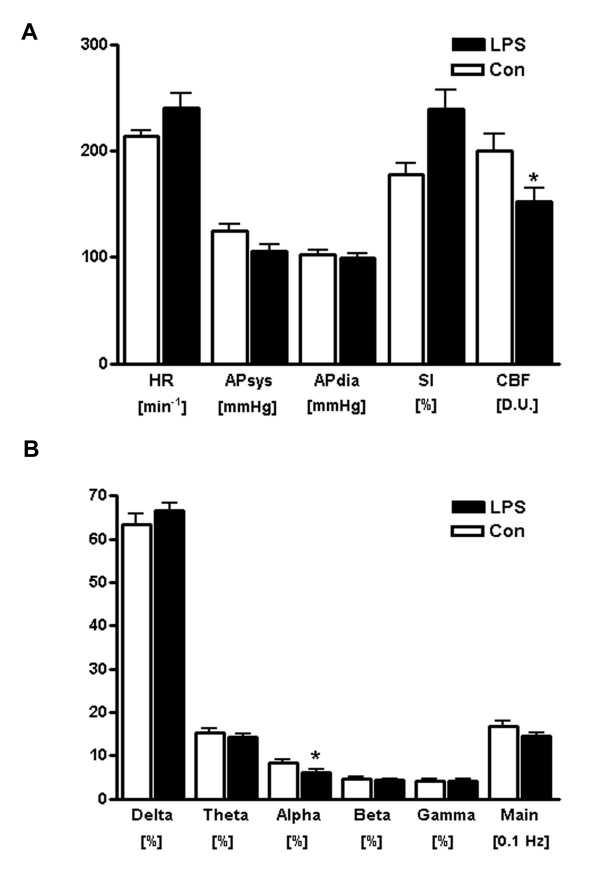
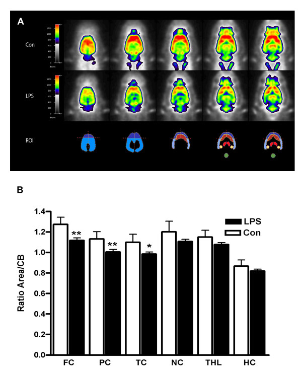
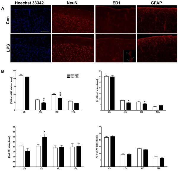
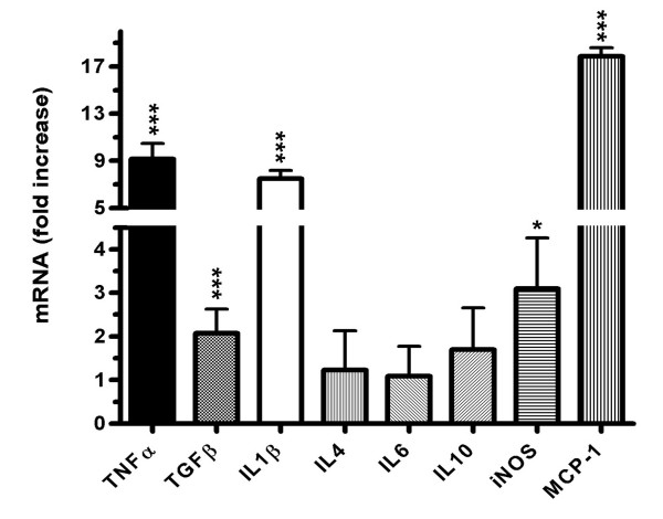
Results: While the systemic hemodynamic did not change significantly, regional cerebral blood flow was decreased in the cortex paralleled by a decrease of alpha activity of the electroencephalography. Cerebral glucose uptake was reduced in all analyzed neocortical areas, but preserved in the caudate nucleus, the hippocampus and the thalamus. Sepsis enhanced the transcription of several pro- and anti-inflammatory cytokines and chemokines including tumor necrosis factor alpha, interleukin-1 beta, transforming growth factor beta, and monocot chemoattractant protein 1 in the cerebrum. Regional analysis of different brain regions revealed an increase in ED1-positive microglia in the cortex, while total and neuronal cell counts decreased in the cortex and the hippocampus.

Conclusion: Together, the present study highlights the complexity of sepsis induced early impairment of neuronal metabolism and activity. Since our model uses techniques that determine parameters relevant to the clinical setting, it might be a useful tool to develop brain specific therapeutic strategies for human septic encephalopathy.


Figures

Figure 1
Figure 1
Changes of hemodynamics and electroencephalography in response to sepsis induction Sample figure title. A, Displayed are heart rate (HR), systolic and diastolic arterial pressure (APsys, APdia), shock index (SI) and cerebral blood flow (CBF) in vehicle-treated (Con) and LPS-treated rats at 24 h after sepsis induction. While the general cardiovascular parameters did not change significantly, CBF was markedly reduced upon LPS exposure. B, Shown is the relative spectral band power and the main spectral frequency determined from EEG recordings in vehicle-(Con) and LPS-treated rats 24 h post induction (mean ± SEM; n = 5 animals/group; two-sided Mann-Whitney test; *p ≤ 0.05, **p ≤ 0.01).
Figure 2
Figure 2
Reduction of cerebral glucose uptake in septic encephalopathy. Shown are five representative transversal [18F]FDG-PET brain slices of rats treated with vehicle (Con, first row) or bacterial lipopolysaccharide (LPS, second row) at 24 h. Corresponding region-of-interest (ROI) masks are displayed below. B, Quantification of [18F]FDG uptake (relative to cerebellar (CB) ROI) in vehicle-treated wild-type (Con) and LPS-treated mice (LPS) at 24 h. Significant differences were detected in frontal cortex (FC), parietal cortex (PC) and temporal cortex (TC). In contrast, evaluation of the caudate nucleus (NC), thalamus (THL) and hippocampus (HC) did not yield significant results (mean ± SEM; n = 5 animals/group; Student's t test; *p ≤ 0.05, **p ≤ 0.01).
Figure 3
Figure 3
Histological analysis of regional LPS induced changes of cell number, micro- and astroglial activation. A, cell number detection by Hoechst 33342 staining and detection of microglial (ED1), neuronal (NeuN), and astroglial (GFAP) immunoreactivity in the cortex, 24 h after intraperitoneal application of LPS (LPS) or vehicle (Con). Results are given as percentage of immunopositive area. The inset shows a 100× magnification of ED1 positive microglial cells located along a cerebral blood vessel. (mean ± SEM; n = 5 animals/group; Student's t test; *p ≤ 0.05, **p ≤ 0.01). B, Quantitative analysis of Hoechst 33342 staining, ED1, NeuN, and GFAP immunostaining in the cortex (Cx), cerebellum (Cb), thalamus (THL), and hippocampus (HC).
Figure 4
Figure 4
Reduction of cerebral glucose uptake in septic encephalopathy. Inflammatory gene transcription was studied using cDNA generated from whole brain lysates 24 h after intraperitoneal administration of either LPS or vehicle. Induction of the indicated pro- and anti-inflammatory cytokines was measured by real-time RT-PCR with RNA samples prepared from vehicle treated controls and LPS treated rats. Triplicate determination was performed for each mRNA sample. Gene-expression levels were normalized with GAPDH for each mRNA preparation, and the n-fold increase in LPS treated animals was calculated by comparison with the result obtained in untreated rats. Results are the mean ± (+SEM) of five animals per group. (Student's t test; *p ≤ 0.05, ***p ≤ 0.001).

References

    1. Pine RW, Wertz MJ, Lennard ES, Dellinger EP, Carrico CJ, Minshew BH. Determinants of Organ Malfunction Or Death in Patients with Intra-Abdominal Sepsis – A Discriminant-Analysis. Archives of Surgery. 1983;118:242–249. - PubMed
    1. Sprung CL, Peduzzi PN, Shatney CH, Schein RMH, Wilson MF, Sheagren JN, et al. Impact of Encephalopathy on Mortality in the Sepsis Syndrome. Critical Care Medicine. 1990;18:801–806. - PubMed
    1. Wilson JX, Young GB. Progress in clinical neurosciences: Sepsis-associated encephalopathy: Evolving concepts. Canadian Journal of Neurological Sciences. 2003;30:98–105. - PubMed
    1. Pickering M, Cumiskey D, O'Connor JJ. Actions of TNF-alpha on glutamatergic synaptic transmission in the central nervous system. Experimental Physiology. 2005;90:663–670. doi: 10.1113/expphysiol.2005.030734. - DOI - PubMed
    1. Young GB, Bolton CF, Archibald YM, Austin TW, Wells GA. The Electroencephalogram in Sepsis-Associated Encephalopathy. Journal of Clinical Neurophysiology. 1992;9:145–152. doi: 10.1097/00004691-199201000-00016. - DOI - PubMed

Publication types

MeSH terms