. 2008 Sep 15;68(18):7264-9.
doi: 10.1158/0008-5472.CAN-08-1365.

Combination therapy with cisplatin and anti-4-1BB: synergistic anticancer effects and amelioration of cisplatin-induced nephrotoxicity

Affiliations

Combination therapy with cisplatin and anti-4-1BB: synergistic anticancer effects and amelioration of cisplatin-induced nephrotoxicity

Young H Kim et al. Cancer Res. .

Abstract

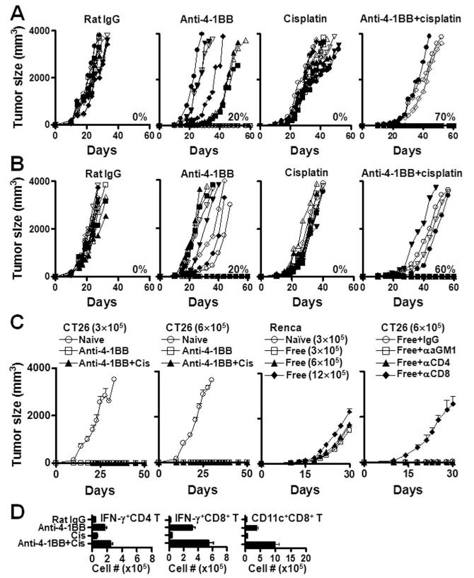
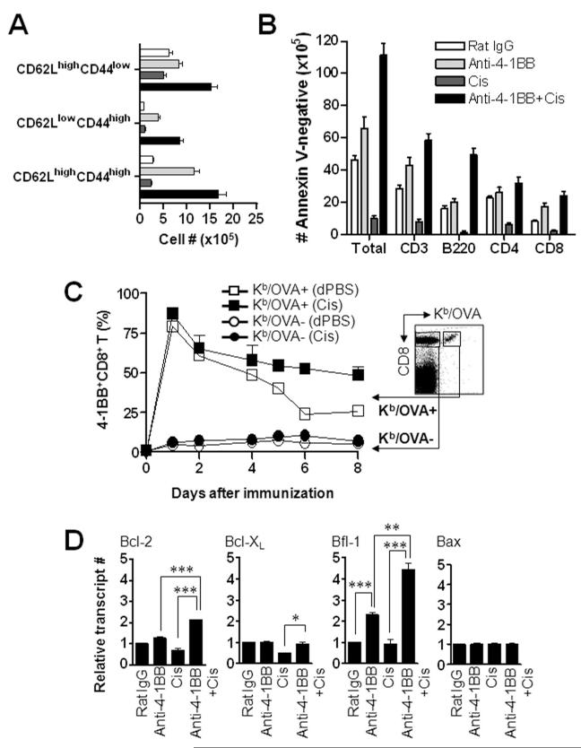
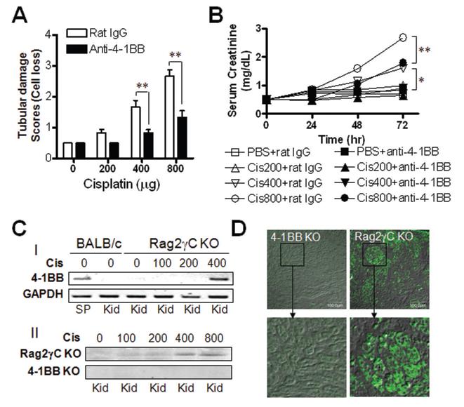
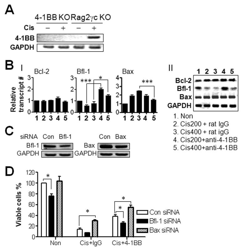
Anti-4-1BB and cisplatin showed synergistic anticancer effects in the CT-26 colon carcinoma model, producing complete regression in >60% of mice with either preventive or therapeutic treatment. The tumor-free mice formed long-lasting CD8(+) T cell-dependent tumor-specific memory. Anti-4-1BB induced rapid repopulation of T and B cells from cisplatin-mediated lymphopenia and differentiation and expansion of IFN-gamma(+)CD11c(+)CD8(+) T cells. Cisplatin facilitated expansion of naïve, effector, and memory CD8(+) T cells; combination therapy produced almost twice as many lymphoid cells as anti-4-1BB alone. Cisplatin increased 4-1BB on antigen-primed T cells and induced 4-1BB de novo on kidney tubular epithelium. Cross-linking of 4-1BB protected the T cells and kidney epithelium from cisplatin-mediated apoptosis by increasing expression of antiapoptotic molecules. Thus, cisplatin-induced 4-1BB provided a mechanism for amelioration of the lymphopenia and nephrotoxicity inherent in cisplatin treatment. We concluded that chemoimmunotherapy with anti-4-1BB and cisplatin is synergistic in tumor killing and prevention of organ-specific toxicity.


Figures

Figure 1
Figure 1
Combination therapy with anti-4-1BB and cisplatin synergistically induces anti-tumor immune responses. (A) BALB/c mice were challenged s.c. with 3 × 10⁵ CT26 colon carcinoma cells, received a single i.p. injection of 200 μg cisplatin, and injected i.p. with 100μg of rat IgG or anti-4-1BB mAb (3E1) every 5 days for five times from PI day 0. (B) Six days after tumor challenge, mice were treated i.p with PBS or 200 μg cisplatin and injected with rat IgG or anti-4-1BB mAb every 5 days for five times. The mice were monitored everyday for tumor growth and survival. (C) Tumor-free mice from the anti-4-1BB- or combination-treated groups were rechallenged s.c. with the indicated number of CT26 or RENCA cells on PI day 100 and monitored for tumor growth. In separated experiment, tumor-free mice on PI day 100 were challenged s.c. with CT26 and treated i.p. with 100μg of anti-asialo-GM1, 400μg of GK1.5, or 400μg of 2.43 mAb to deplete NK, CD4+ T, or CD8+ T cells every 5 days for five times. (D) On PI day 17, single cell suspensions of TDLNs were stained with PE-anti-CD11c and PE-Cy5-anti-CD8. TDLN cells were also stained with FITC-anti-CD4 or -anti-CD8, and further intracellularly stained with PE-anti-IFN-γ, and subsequently analyzed using FACscan (BD Biosciences). Absolute numbers were calculated by multiplying percentages measured by flow cytometry by total numbers of viable cells. Each group contained 10 mice (A-B) or 5 mice (C-D). The results are representative of two independent experiments. All plotted data are means ± SD.
Figure 2
Figure 2
Treatment with anti-4-1BB rescues lymphocytes from cisplatin-mediated apoptosis. Tumor-challenged BALB/c mice were injected i.p. with rat IgG or anti-4-1BB mAb as well as single injection of cisplatin on PI day 0 as described above. On PI day 17, single cell suspensions of TDLNs were stained with FITC-anti-CD62L, PE-anti-CD44, and PE-Cy5-anti-CD8 (A). (B) Five days after the treatment, TDLN cells were counted and stained with FITC-anti-CD3, -anti-B220, -anti-CD4 or -anti-CD8, and further stained with PE-anti-Annexin-V to examine the apoptotic cells. Samples were subsequently analyzed using FACscan and absolute numbers of annexin V-negative cells were calculated as described above. (C) To create a system that determine the 4-1BB expression on both Ag-specific and Ag-nonspecific CD8+ T cells simultaneously, OVA-specific CD8+ T cells were isolated from OT-I mice and adoptively transferred into C57BL/6 mice (5 × 10⁶ cells per mouse). The mice were immunized s.c. with 10 μg of OVA in incomplete Freund’s adjuvant (IFA), and injected i.p. with 200 μg of cisplatin (filled symbol) or PBS (open symbol) as a control. Draining lymph node (DLN) cells were stained with FITC-anti-4-1BB, PE-Kb/OVA tetramer, and PE-Cy5-anti-CD8 on the indicated days. Data dictated the percentage of 4-1BB+CD8+ T cells in gated Kb/OVA tetramer-negative or Kb/OVA tetramer-positive CD8+ T cells. (D) Total RNA was extracted from CD8+ T cells of four different groups of mice on PI day 5 and first-strand cDNA was synthesized with 0.5 μg total RNA. SYBR Green I-based real-time quantitative PCR was carried out on a continuous fluorescence detection system (Opticon DNA Engine, MJ Research Inc, Waltham, MA). Relative gene expression was determined using the comparative CT method and normalized to the housekeeping gene GAPDH. To simply represent Bcl-2, Bcl-XL, Bfl-1, and Bax expression, the gene expression data normalized for GAPDH are shown as the fold increase compared to levels in the rat IgG-treated group. Each group contained three mice and the results are representative of two independent experiments. All plotted data are means ± SD (*, P < 0.05; **, P < 0.01; ***, P < 0.001).
Figure 3
Figure 3
4-1BB triggering ameliorates cisplatin-mediated nephrotoxicity. BALB/c mice were injected i.p. with various doses of cisplatin and simultaneously injected with rat IgG or anti-4-1BB mAb. (A) Semiquantification of mouse renal tubular damage. Mouse kidneys at 72hrs after cisplatin injection were embedded in paraffin, cut into 4 mm sections, and stained with haematoxylin-eosin (H&E). Renal tubular damage was assessed using a semiquantitative scale. A pathologist who was blinded to the experiments scored the degree of tubular damage. The magnitude of tubular epithelial cell necrosis and loss was scored on the basis of the percentage of affected tubules in a high-power field under a light microscope, as follows: 0, normal; 0.5, < 10%; 1, 10 to 25%; 2, 25 to 50%; 3, 50 to 75%; and 4, > 75%. (B) Blood samples were obtained from before (0 hour) and at 24, 48, and 72 h after treatment with cisplatin and/or anti-4-1BB mAb. Serum creatinine was measured as a marker of renal dysfunction using a Roche Cobas Rara automated system (Roche, Nutley, NJ) with a Creatinine kit (R&D Systems, Minneapolis, MN). (C) 4-1BB expression in renal tubular epithelial cells. I. Rag2γc- or 4-1BB-deficient mice received a single injection of 100, 200 or 400 μg cisplatin. RT-PCR for 4-1BB was performed with cDNA of kidney cells 24 hr after cisplatin administration. II. 4-1BB proteins were detected in kidney samples by performing western blotting with anti-4-1BB mAb (3E1 clone). (D) For anti-4-1BB immunostaining of the kidney, frozen sections (4 μm thickness) were prepared from kidney 24 h after cisplatin administration and stained with FITC-conjugated anti-m4-1BB mAb. Sections were viewed and photographed using a laser-scanning confocal fluorescence microscope system (Fluoview FV500 Olympus, Center Valley, PA; original magnification ×10). Each group contained three mice and the results are representative of three independent experiments. All plotted data are means ± SD (*, P < 0.05; **, P < 0.01; ***, P < 0.001).
Figure 4
Figure 4
4-1BB triggering enhances survival of kidney epithelium in vivo. (A) Kidney epithelial cells from 4-1BB KO BALB/c or Rag2γc KO mice were cultured in vitro for 7 days and treated with 2 μg/ml cisplatin or PBS for 24hrs. RT-PCR was performed with cDNA of the cultured cells to determine 4-1BB transcripts. (B) Rag2γc KO mice were injected i.p. with indicated doses of cisplatin and also single-injected with 100μg of rat IgG or anti-4-1BB mAb. Complementary DNA was synthesized with total RNA from kidney 72 hrs after cisplatin injection. Real-time PCR (I) and RT-PCR (II) were performed to assess Bcl-2, Bfl-1, and Bax expression as describe above. Relative gene expression was normalized to GAPDH and shown as the fold increase compared to that of rat IgG-treated group. (C, D) Kidney epithelial cells from Rag2γc DKO or 4-1BB KO mice were transfected with either gene-specific siRNA against Bfl-1, Bax or nontargeting control siRNA according to manufacturer’s instruction (Santa Cruz Biotechology). (C) Decrease in Bfl-1 or Bax mRNA upon corresponding siRNA transfection was determined by RT-PCR. (D) Cells were treated with 2 μg/ml cisplatin and/or 10 μg/ml of rat IgG or anti-4-1BB, and the absolute numbers of viable cells were determined at the indicated time points. Each group contained three mice and the results are representative of three independent experiments. All plotted data are means ± SD (*, P < 0.05; **, P < 0.01; ***, P < 0.001).

Similar articles

Cited by

References

    1. Nowak AK, Lake RA, Marzo AL, et al. Induction of tumor cell apoptosis in vivo increases tumor antigen cross-presentation, cross-priming rather than cross-tolerizing host tumor-specific CD8 T cells. J Immunol. 2003;170(10):4905–13. - PubMed
    1. Nowak AK, Robinson BW, Lake RA. Synergy between chemotherapy and immunotherapy in the treatment of established murine solid tumors. Cancer Res. 2003;63(15):4490–6. - PubMed
    1. Bergmann-Leitner ES, Abrams SI. Treatment of human colon carcinoma cell lines with anti-neoplastic agents enhances their lytic sensitivity to antigen-specific CD8+ cytotoxic T lymphocytes. Cancer Immunol Immunother. 2001;50(9):445–55. - PMC - PubMed
    1. Dudley ME, Wunderlich JR, Robbins PF, et al. Cancer regression and autoimmunity in patients after clonal repopulation with antitumor lymphocytes. Science. 2002;298(5594):850–4. - PMC - PubMed
    1. Ghiringhelli F, Larmonier N, Schmitt E, et al. CD4+CD25+ regulatory T cells suppress tumor immunity but are sensitive to cyclophosphamide which allows immunotherapy of established tumors to be curative. Eur J Immunol. 2004;34(2):336–44. - PubMed

Publication types

MeSH terms