. 2008 Sep 22;182(6):1217-30.
doi: 10.1083/jcb.200712086. Epub 2008 Sep 15.

EPB41L5 functions to post-transcriptionally regulate cadherin and integrin during epithelial-mesenchymal transition

Affiliations

EPB41L5 functions to post-transcriptionally regulate cadherin and integrin during epithelial-mesenchymal transition

Mariko Hirano et al. J Cell Biol. .

Abstract

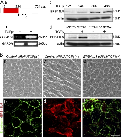
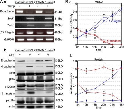
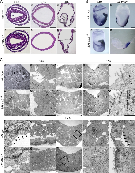
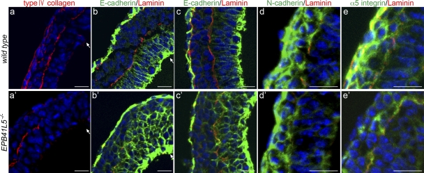
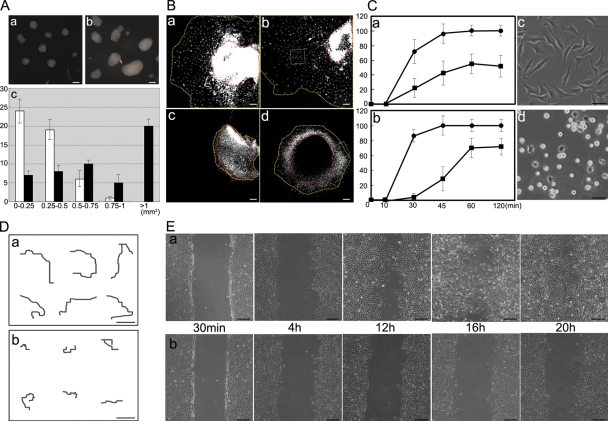
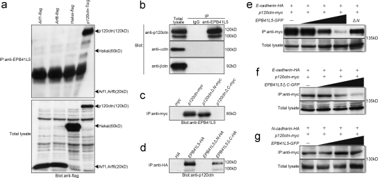
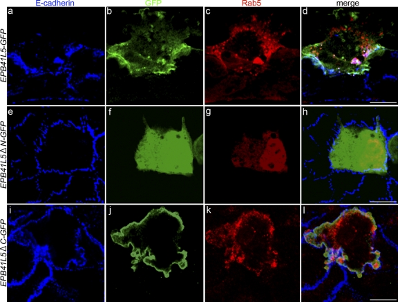
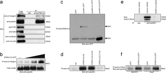
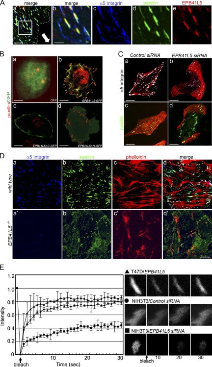
EPB41L5 belongs to the band 4.1 superfamily. We investigate here the involvement of EPB41L5 in epithelial-mesenchymal transition (EMT) during mouse gastrulation. EPB41L5 expression is induced during TGFbeta-stimulated EMT, whereas silencing of EPB41L5 by siRNA inhibits this transition. In EPB41L5 mutants, cell-cell adhesion is enhanced, and EMT is greatly impaired during gastrulation. Moreover, cell attachment, spreading, and mobility are greatly reduced by EPB41L5 deficiency. Gene transcription regulation during EMT occurs normally at the mRNA level; EPB41L5 siRNA does not affect either the decrease in E-cadherin or the increase in integrin expression. However, at the protein level, the decrease in E-cadherin and increase in integrin are inhibited in both EPB41L5 siRNA-treated NMuMG cells and mutant mesoderm. We find that EPB41L5 binds p120ctn through its N-terminal FERM domain, inhibiting p120ctn-E-cadherin binding. EPB41L5 overexpression causes E-cadherin relocalization into Rab5-positive vesicles in epithelial cells. At the same time, EPB41L5 binds to paxillin through its C terminus, enhancing integrin/paxillin association, thereby stimulating focal adhesion formation.


Figures

Figure 1.
Figure 1.
EPB41L5 functions in EMT of NMuMG cells by TGFβ. (A) EPB41L5 expression in NMuMG cells. (a) Schematic representation of EPB41L5. Red represents FERM domain. An arrow indicates the point of truncation in the analyses of Fig. 6 (d–f), Fig. 7 (e–l), Fig. 8 (d), and Fig. 9 (Bc and d). The underline indicates the location of the sequences used as an antigen to raise an antibody. Arrowheads (F, R) indicate the locations of primers for RT-PCR analyses in b. (b and c) The increase in EPB41L5 expression with the TGFβ treatment assayed by RT-PCR (b) at 48 h and Western blotting (c) at the indicated time. (d) Effect of EPB41L5 siRNA treatment on the EPB41L5 protein induction by TGFβ. The cells were transfected with control or EPB41L5 siRNA and cultured in the absence or presence of TGFβ for 48 h. (B) Inhibition of TGFβ-induced EMT of NMuMG cells by EPB41L5 siRNA. EPB41L5 (red) is scarce in the confluent epithelial NMuMG cells in the absence of TGFβ; E-cadherin (green) is abundant at cell–cell contact sites (a and b). In “mesenchymal” cells induced by TGFβ, EPB41L5 is abundantly present with no E-cadherin expression (c and d). EPB41L5 siRNA inhibits the transformation into “mesenchyme” by TGFβ (e), retaining E-cadherin at cell–cell contact sites (f). Bars, 20 μm.
Figure 2.
Figure 2.
Changes in E-cadherin and β1-integrin expression by TGFβ and/or EPB41L5 siRNA treatment of NMuMG cells. (A) RT-PCR (a) and Western blot (b) analysis. Cells transfected with control or EPB41L5 siRNA were plated at 104/cm2 and cultured in the absence (−) or presence (+) of TGFβ for 48 h, and the expression of each molecule indicated was determined. (B) Quantitation of E-cadherin (red lines) and β1-integrin (blue lines) expression by RT-PCR (a) and Western blotting (b) at the indicated time after plating. Circles represent the cells transfected with control siRNA, and squares those with EPB41L5 siRNA. Ordinates give the relative intensity to GAPDH (a) or actin (b) expression, respectively, determined with ImageJ software. The experiment was repeated three times.
Figure 3.
Figure 3.
EPB41L5 mutant phenotype. (A) Histological views of wild-type (a–c) and mutant (a′–c′) embryos at E6.5 (a and a′), E7.5 (b and b′), and E8.5 (c and c′), respectively. (a, a′, b, and b′) Horizontal sections. (c and c′) Sagittal sections. Anterior is to the left. Bars, 100 μm. (B) Whole-mount RNA in situ hybridization views of wild-type (a and b) and mutant (a′ and b′) embryos at E7.5 for Snail (a and a′) and Brachury (b and b′) expression. Lateral views; anterior is to the left. Bars, 100 μm. (C) Electron microscopic views of EPB41L5 mutant embryos. (a–j) Views in wild-type. (a′–j′) Views in EPB41L5 mutant embryos. (a and a′) Views at E6.5 primitive streak. (b and b′) Enlarged views of the regions boxed in a and a′, respectively. (c and c′) Views of the lateral region of E6.5 embryos. (d and d′) Views of the anterior region of E7.5 embryos. (e–f′) Enlarged views of the regions boxed in d and d′. (g and g′) Views of laterally invaginated E7.5 mesoderm cells. (h and h′) Views of cells invaginating at E7.5 primitive streak. (I and i′) Enlarged views of the regions boxed in (h and h′). (j and j′) Views of E7.5 endoderm cells. Dotted lines in d and d′ outline individual cells in the anterior-most ectoderm layers; these cells are ectodermal, as suggested by the location of residual basement membrane components and E-cadherin expression shown in Fig. 4, b′ and c′. Arrows in f and j indicate basement membranes. In e and e′, TJ is tight junction; AJ, adherens junction; De, desmosome. Bars, 5 μm.
Figure 4.
Figure 4.
Marker analyses of epithelial mesenchyme transition in EPB41L5 mutant gastrula. (a–e) Wild-type and (a′–e′) mutant embryos at E7.5. Red indicates type IV collagen staining in a and a′ and laminin staining in b–e′ for basement membranes, and blue DAPI staining for nucleus in all figures. (b–c′) E-cadherin staining (green) at the primitive streak region (b and b′) and lateral region (c and c′). (d and d′) N-cadherin. (e and e′) α5-integrin staining (green). Arrows indicate invagination sites. (c′) Ectodermal nature of the multi-layered cells by the locations of residual laminin-positive basement membranes and E-cadherin expression (compare Fig. 3 Cd′). Bars, 20 μm.
Figure 5.
Figure 5.
Roles of EPB41L5 in cell–cell and cell–substrate interaction. (A) Cell aggregation assay. NMuMG cells were treated with TGFβ for 48 h and transfected with (a) control or (b) EPB41L5 siRNA. After 12 h, cells were trypsinized and cultured in suspension for 4 h as described previously (Takeichi, 1977). (a and b) Examples of cell aggregates. (c) Size distribution of the aggregates; the size is represented as the product of the major and minor axes. 50 aggregates were counted, and their frequency in the size range indicated at the abscissa is given in the ordinate for control (white columns) and EPB41L5 (black columns) siRNA-treated cells. Bars, 200 μm. (B) Effects of anti–E-cadherin antibody on mobility of EPB41L5 mutant primitive streak cells. Primitive streak was dissected from wild-type (a and b) or mutant (c and d) E7.5 embryos and cultured with control DMSO (a and c) or rat anti–mouse E-cadherin antibody (b and d) for 2 d as described previously (Zohn et al., 2006). Red lines indicate the peripheries of primitive streak cell mass and yellow lines the forefront of migrating cells. See Fig. 9 D for the boxes in b and d. Bars, 200 μm. (C) Cell attachment assay to fibronectin-coated substrates. (a) The assay with wild-type (•) and EPB41L5 mutant (▪) cells dispersed by trypsinization and pipetting from E7.5 whole embryos; (b), the assay with NIH3T3 cells transfected with control (•) or EPB41L5 (▪) siRNA. (c and d) Spreading of NIH3T3 cells transfected with control (c) or EPB41L5 (d) siRNA after 120 min culture. NIH3T3 cells express EPB41L5 at the comparable level to that in TGFβ-treated NMuMG cells shown in Fig. 1 A. Bars, 100 μm. (D) Cell movement on the fibronectin-coated substrates. NIH3T3 cells transfected with control (a) or EPB41L5 (b) siRNA were plated sparsely, and after 8 h the movement of individual cells was traced by time-lapse cinematography for 12 h. Typical examples of the tracing are shown on six cells, respectively. Bars, 100 μm. (E) Wound-healing assay on the fibronectin-coated substrates. (a and b) NIH3T3 cells transfected with control (a) or EPB41L5 (b) siRNA were plated at a high cell density, and the monolayers were scratched 12 h after plating and cultured for the indicated times. Bars, 200 μm. Similar observations on cell attachment, spreading, and mobility obtained with TGFβ-treated NMuM G cells are presented in Fig. S4.
Figure 6.
Figure 6.
EPB41L5 interaction with E-cadherin–associated adhesion components. (a) EPB41L5 binding to Arf1, Arf6, Hakai, and p120 catenin. Each expression vector harboring Arf1, Arf6, Hakai, or p120 catenin (p120ctn) linked with flag was transfected into Cos7 cells 12 h after plating, and the cells were harvested 24 h later. The bottom panel gives the Western blotting for the expression of each transgene in total cell lysates of respective transfectants. In the top panel the lysates were immunoprecipitated with the anti-EPB41L5 antibody, and the presence or absence of each molecule in the immunoprecipitates was blotted with anti-flag antibody. (b) EPB41L5 binding to p120ctn, α-catenin (αctn) and β-catenin (βctn). The left panel gives the expression level of each catenin in TGFβ-treated NMuMG cells. In the other two lanes the cell lysates were immunoprecipitated with rabbit IgG (middle) or rabbit anti-EPB41L5 antibody (right), and the presence of each catenin in the precipitates was blotted with the antibody against each catenin. (c) EPB41L5 binding to p120 catenin. Myc, myc-linked full-length p120ctn, myc-linked p120ctn that lacks N-terminal (ΔN), or myc-linked p120ctn lacking C-terminal armadillo repeats (ΔC) was transfected into Cos7 cells as described in panel a, cell lysates were immunoprecipitated with anti-myc antibody, and the presence or absence of EPB41L5 in the precipitates was blotted with anti-EPB41L5 antibody. (d) p120 catenin binding to EPB41L5. HA, HA-linked full-length EPB41L5, HA-linked EPB41L5 that lacks N-terminal FERM domain (ΔN), or HA-linked EPB41L5 lacking C-terminal (ΔC) was transfected into Cos7 cells, cell lysates were immunoprecipitated with anti-HA antibody, and the presence or absence of p120ctn in the precipitates was blotted with anti-p120ctn antibody. See Fig. 1 A for the site of the EPB41L5 truncation. (e) Effects of EPB41L5 on the binding between E-cadherin and p120ctn. 1 μg HA-linked E-cadherin and myc-linked p120ctn were cotransfected with 0, 1, 2.5, or 5 μg GFP-linked full-length EPB41L5 or with 5 μg GFP-linked EPB41L5 that lacks the N-terminal (ΔN). The bottom panel gives the amount of E-cadherin in total cell lysates. The lysates were immunoprecipitated with anti-myc antibody for p120ctn and the amount of E-cadherin in the precipitates was blotted with anti-HA antibody. (f) Effects of EPB41L5 that lacks C-terminal but retains N-terminal FERM domain on the binding between E-cadherin and p120ctn. (g) Effects of EPB41L5 on the binding between N-cadherin and p120ctn. The binding assay in f and g was conducted as described in panel e.
Figure 7.
Figure 7.
Effects of EPB41L5 on E-cadherin and Rab5 distribution. GFP-linked full-length EPB41L5 (a–d), GFP-linked EPB41L5 that lacks N-terminal FERM domain (e–h), or GFP-linked EPB41L5 that lacks C terminus (i–l) was co-transfected with myc-linked Rab5 (to detect endosomes) into epithelial NMuMG cells. (a, e, and i) Endogenous E-cadherin staining (blue), (b, f, and j) GFP staining for EPB41L5 localization (green), (c, g, and k) myc staining for Rab5-positive endosomes (red), and (d, h, and l) merged views. Confluent epithelial NMuMG cells, in the absence of TGFβ, uptake little DNA, and the rare cells that took up DNAs are marked by green and red among most cells that did not (blue). myc-Rab5 itself has no effect on E-cadherin distribution (e–h) (Palacios et al., 2005). In the NMuMG cells that took up full-length EPB41L5 (a–d) or EPB41L5 that lacks C terminus (i–l), the number of E-cadherin–positive vesicles is increased in cytoplasm. The E-cadherin–positive vesicles coincided with Rab5-positive endosomes (d and l); Rab5-positive vesicles are also enhanced. EPB41L5 that lacks N-terminal FERM domain (e–h) does not increase cytoplasmic E-cadherin–positive vesicles. In each transfection more than 30 GFP-positive cells were examined, all of the cells showing the pattern of EPB41L5, E-cadherin and Rab5 localization shown here. Bars, 30 μm. Similar observations obtained with T47D cells are presented in Fig. S5 A.
Figure 8.
Figure 8.
EPB41L5 interaction with integrin-associated adhesion components. (a) EPB41L5 binding to integrin-associated adhesion components in TGFβ-treated NMuMG cells. The amount of each component in the total cell lysates is given in the left panel. In the other two lanes the cell lysates were immunoprecipitated with rabbit IgG (middle) or rabbit anti-EPB41L5 antibody (right), and the presence of each component in the precipitates was blotted with the antibody against each molecule indicated at left. (b) Effects of EPB41L5 on the binding between β1-integrin and paxillin. 0, 1, 2.5, or 5 μg GFP-linked full-length EPB41L5 was transfected into Cos7 cells, and the binding assay was performed 24 h later. The bottom panel gives the amount of paxillin in total cell lysates. In the top panel the lysates were immunoprecipitated with anti-β1-integrin antibody, and the amount of paxillin in the precipitates was blotted with anti-paxillin antibody. (c) EPB41L5 binding to paxillin. GFP, GFP-linked full-length paxillin, GFP-linked paxillin that lacks C-terminal (ΔC), or GFP-linked paxillin lacking N-terminal (ΔN) was transfected into Cos7 cells as described in Fig. 6 a, cell lysates were immunoprecipitated with anti-EPB41L5 antibody, and the presence or absence of paxillin in the precipitates was blotted with anti-GFP antibody. (d) Paxillin binding to EPB41L5. HA, HA-linked full-length EPB41L5, HA-linked EPB41L5 that lacks N-terminal FARM domain (ΔN), or HA-linked EPB41L5 lacking C terminus (ΔC) was transfected into Cos7 cells, cell lysates were immunoprecipitated with anti-HA antibody, and the presence or absence of paxillin in the precipitates was blotted with anti-paxillin antibody. See Fig. 1 A for the site of the EPB41L5 truncation. (e) Effects of EPB41L5 siRNA on p120ctn binding to paxillin. NIH3T3 cells were transfected with control or EPB41L5 siRNA and harvested 24 h later; cell lysates were immunoprecipitated with rabbit IgG or rabbit anti–mouse paxillin antibody, and the amount of p120ctn in the precipitates was blotted with anti-p120ctn antibody. (f) Effects of EPB41L5 overexpression on p120ctn binding to paxillin. NIH3T3 or Cos7 cells were transfected with GFP or GFP-linked EPB41L5; cell lysates were immunoprecipitated with anti-p120ctn antibody, and paxillin in the precipitates was blotted with anti-paxillin antibody.
Figure 9.
Figure 9.
Effects of EPB41L5 on focal adhesion formation and turnover. (A) Colocalization of EPB41L5 with α5-integrin and paxillin at the leading edge of TGFβ-treated mesenchymal NMuMG cells. Monolayered NIH3T3 cells were scratched and cultured for 12 h (Fig. 5 Ea); the dotted line indicates the cell periphery, and the direction of the cell movement is indicated by an arrow in panel a. Bars, 10 μm. (b–e) Enlarged views of the boxed area in panel a. (a and b) Merged views of (c) α5-integrin (blue), (d) paxillin (green), and (e) EPB41L5 (red) localization. Bars, 2 μm. Endogenous paxillin and EPB41L5 localization in TGFβ-treated mesenchymal NMuMG cells is also given in Fig. S5 B. (B) Increase in focal adhesions by EPB41L5 expression in T47D cells. GFP (a), GFP-linked full-length EPB41L5 (b), GFP-linked EPB41L5 that lacks C-terminal paxillin binding domain (ΔC) (c), or GFP-linked EPB41L5 lacking N-terminal FERM domain (ΔN) (d) was transfected into T47D cells that do not express EPB41L5. The cells are stained for GFP (green) and endogenous paxillin (red). Quantification of paxillin-positive structures is given in Fig. S5 C. Bars, 30 μm. (C) Disappearance of focal adhesions in NIH3T3 cells by EPB41L5 siRNA. The cells were transfected with control (a and c) or EPB41L5 siRNA (b and d) and stained for actin (red) and α5-integrin (a and b; white) or paxillin (c and d; green). Bars, 30 μm. (D) Distribution of focal adhesions in wild-type (a–d) and EPB41L5 mutant (a′–d′) cells. α5-Integrin (a and a′), paxillin (b and b′), and actin (c and c′) expression and merged views (d and d′) are shown on the cells that crept out from the primitive streak cell mass as boxed in Fig. 5, Bb and d. Quantification of paxillin- and α5-integrin–positive structures is given in Fig. S5 D. Bars, 50 μm. (E) FRAP assay. FRAP analysis was conducted with paxillin-GFP expressing T47D cells transfected with EPB41L5 (▴) or with paxillin-GFP expressing NIH3T3 cells transfected with control (•) or EPB41L5 siRNA(▪). Right panels show examples of fluorescence recovery after photobleaching in the T47D cell overexpressing EPB41L5 (top), control NIH3T3 cell (middle), and NIH3T3 cells treated with EPB41L5 siRNA (bottom).

Similar articles

Cited by

References

    1. Aono, S., S. Nakagawa, A.B. Reynolds, and M. Takeichi. 1999. p120(ctn) acts an inhibitory regulator of cadherin function in colon carcinoma cells. J. Cell Biol. 145:551–562. - PMC - PubMed
    1. Batlle, E., E. Sancho, C. Franci, D. Dominguez, M. Monfar, J. Baulida, and A. Garcia de Herreros. 2000. The transcription factor snail is a repressor of E-cadherin gene expression in epithelial tumour cells. Nat. Cell Biol. 2:84–89. - PubMed
    1. Cano, A., M.A. Perez-Moreno, I. Rodrigo, A. Locascio, M.J. Blanco, M.G. del Barrio, F. Portillo, and M.A. Nieto. 2000. The transcription factor snail controls epithelial-mesenchymal transitions by repressing E-cadherin expression. Nat. Cell Biol. 2:76–83. - PubMed
    1. Carver, E.A., R. Jiang, Y. Lan, K.F. Oram, and T. Gridley. 2001. The mouse snail gene encodes a key regulator of the epithelial-mesenchymal transition. Mol. Cell. Biol. 21:8184–8188. - PMC - PubMed
    1. Davis, M.A., R.C. Ireton, and A.B. Reynolds. 2003. A core function for p120-catenin in cadherin turnover. J. Cell Biol. 163:525–534. - PMC - PubMed

Publication types

MeSH terms