. 2008 Sep 29;205(10):2369-79.
doi: 10.1084/jem.20080897. Epub 2008 Sep 15.

The tight junction protein CAR regulates cardiac conduction and cell-cell communication

Affiliations

The tight junction protein CAR regulates cardiac conduction and cell-cell communication

Ulrike Lisewski et al. J Exp Med. .

Abstract

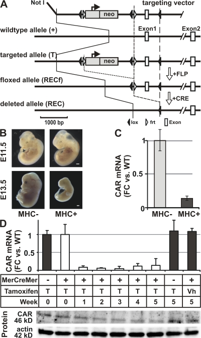
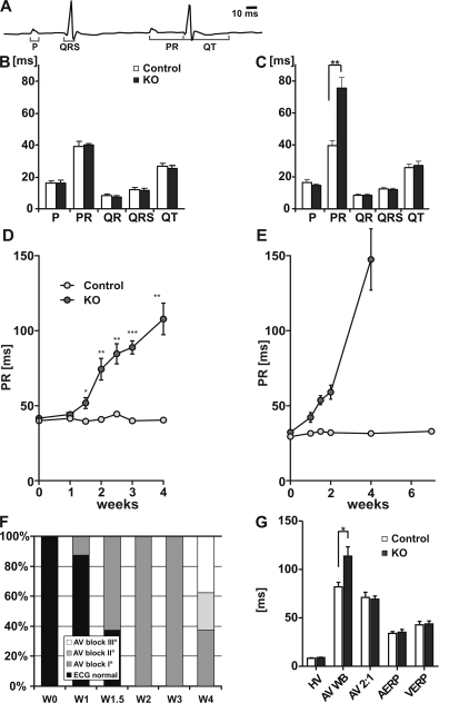
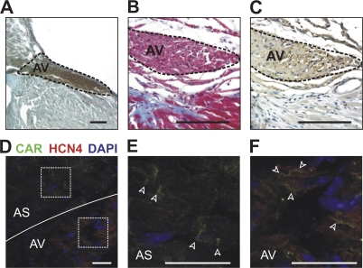
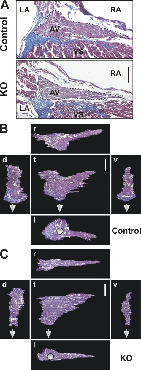
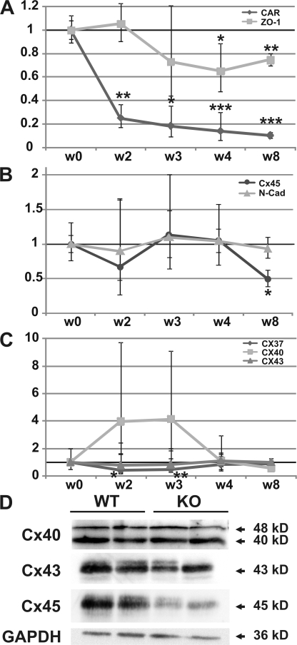
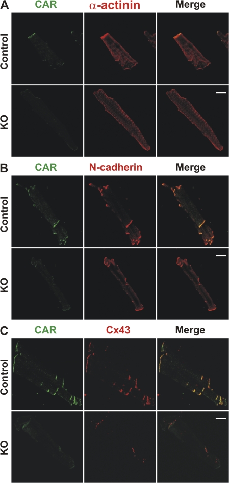
The Coxsackievirus-adenovirus receptor (CAR) is known for its role in virus uptake and as a protein of the tight junction. It is predominantly expressed in the developing brain and heart and reinduced upon cardiac remodeling in heart disease. So far, the physiological functions of CAR in the adult heart are largely unknown. We have generated a heart-specific inducible CAR knockout (KO) and found impaired electrical conduction between atrium and ventricle that increased with progressive loss of CAR. The underlying mechanism relates to the cross talk of tight and gap junctions with altered expression and localization of connexins that affect communication between CAR KO cardiomyocytes. Our results indicate that CAR is not only relevant for virus uptake and cardiac remodeling but also has a previously unknown function in the propagation of excitation from the atrium to the ventricle that could explain the association of arrhythmia and Coxsackievirus infection of the heart.


Figures

Figure 1.
Figure 1.
Cardiac-specific CAR KOs. (A) Targeting strategy. Exon 1, which contains the translation start, was replaced with the floxed exon 1 and the FRT-flanked neo cassette. The neo cassette was subsequently removed by germline expression of the FLP recombinase. We generated a heart-specific and an inducible heart-specific CAR KO using the MHC promoter to express the cre recombinase (MHCcre) or the MerCreMer fusion protein (MCMcre), respectively. The latter is activated by the injection of tamoxifen (30 mg per kilogram body weight per day for 2 wk with five injections per week). Bar, 1,000 bp. (B) The heart-specific KO (MHC+) was unaffected through E11.5 with subsequent failure to thrive and death by day 13.5 of gestation (E13.5). Bar, 500 μm. (C) At E11.5, CAR mRNA levels were reduced to 10% of WT levels (FC, fold change). (D) The tamoxifen-inducible heart-specific CAR KO animals can be induced to excise the CAR exon 1. After 1 wk of tamoxifen injections, CAR mRNA expression dropped to <10% in KO animals, and protein levels, as determined by Western blot (CAR compared with actin), followed at 2 wk as compared with two sets of controls, vehicle (Vh)-injected MCM+ or Tamoxifen-treated MCM− animals. Error bars show SD.
Figure 2.
Figure 2.
Electrical conduction from atrium to ventricle is impaired in CAR KO hearts. (A) Annotated ECG curve. The PR interval corresponds to the time between atrial and ventricular depolarization. (B and C) Quantification of the ECG changes in KO and control animals before (B) and after (C) the 2-wk treatment with tamoxifen (n = 8 per group; D) revealed an increased PR interval in induced KO animals (n = 8 per group). (E) This finding was reproduced by telemetry to exclude an effect of the anesthesia (n = 3 per group). After week 6, all KO animals showed a complete block so that a PR interval could not be derived. (F) The increased PR interval is the ECG correlate of an AV conduction block. Grading the conduction defect in normal AV conduction versus AV block I to III shows the increase in conduction defects over time with third degree AV blocks at week 4 after induction of the KO. (G) As determined by EPS catheter at week 4 after induction, the HV interval, AV node 2:1 conduction capacity (AV 2:1), atrial effective refractory period (AERP), and ventricular effective refractory period (VERP) were unchanged between genotypes, which would exclude an affliction downstream of the HIS bundle. The only significant change was detected in the interval at which electrical propagation from atrium to ventricle was skipped for the first time (Wenckebach Periodicity [AV-WB]) with a significant increase in the KO heart. *, P ≤ 0.05; **, P ≤ 0.01; ***, P ≤ 0.001. Error bars show SEM. W, week.
Figure 3.
Figure 3.
CAR expression in the AV node. (A) The AV node was localized by staining for AChE enzyme activity. (B) Trichrome staining revealed the fibrous body (blue), an anatomical landmark which facilitates localization of the juxtaposed AV node. (C) The identification was confirmed in serial sections by anti-ChAT immunohistochemistry. Bars, 100 μm. (D) Expression of CAR in the AV node was detected by staining with anti-CAR (green) and anti-HCN4 (red) antibodies. The squares indicate the region that is magnified in E and F. (E) CAR is localized in the intercalated disc (arrowheads) within the atrial septum (AS). (F) In cells of the AV node, CAR is distributed along the membrane (arrowheads). Bars, 25 μm.
Figure 4.
Figure 4.
Reduced size of the AV node in the adult CAR KO heart. (A) Masson's Trichrome staining of a KO and control heart at 8 wk after induction shows connective tissue of the valves in blue to separate the AV node area and ventricular septum (VS). To facilitate orientation, left and right atria (LA and RA, respectively) are indicated. The largest AV node area of a series of serial section is slightly reduced in the KO heart compared with the control. (B and C) Three dimensional reconstruction of the AV node from control (B) and CAR KO (C) animals was used to obtain projections in different plains (t, top; l, left; r, right; d, dorsal; v, ventral). The reduced thickness of the AV node results in a volume that corresponds to ∼2/3 of the volume of the control AV node (n = 4). The continuation of the HIS bundle is indicated by white arrows. Bars, 100 μm.
Figure 5.
Figure 5.
Altered cell–cell communication in the CAR KO heart. (A) After injection of fluorescein, the dye is passed to neighboring cells. Representative cells from KO and control cardiac slices from weeks 2–4 after induction of the KO are shown. The increased longitudinal and lateral coupling is most prominent in cardiac slices at the 4-wk time point. (B) Quantification of >200 injections representing 14 independent hearts documents the significantly increased coupling from week 3 after induction of the KO. The area at the end of the 4-min injection period was used to compare cell–cell communication at the indicated times. For each time point, the number of injections and number of animals is provided (injections/animals). **, P ≤ 0.01; ***, P ≤ 0.001, Mann-Whitney test. Error bars show SEM. Bar, 100 μm.
Figure 6.
Figure 6.
Altered expression of the cell–cell contact proteins in CAR KO hearts. (A) Expression of the CAR and its adaptor protein ZO-1 were reduced significantly from weeks 2 and 4, respectively, after induction of the KO. (B) The composition of the GAP junction was altered as a result of differential expression of Cx37 (transiently reduced) and Cx40 (transiently increased, albeit not significantly). Expression of Cx43 was unchanged. (C) Cx45 was the only connexin with RNA levels significantly altered late in the progression of the phenotype (reduced from >8 wk). The adherens junction protein N-cadherin was unchanged. All expression data were normalized to GAPDH and levels at week 0 (before induction) were set to 100%. Fold change is shown on the y-axis. n = 3 per group. Error bars show SD. (D) Cardiac expression of Cx40, Cx43, and Cx45 was confirmed on the protein level with down-regulation of Cx43 and Cx45, but not Cx40, in animals at >8 wk after induction of the phenotype. GAPDH was used as a loading control. After normalization to GAPDH, Cx45 levels were reduced by 45% (P = 0.012), Cx 43 levels by 43% (P = 0.015), and Cx40 levels were not changed significantly (n = 4). *, P ≤ 0.05; **, P ≤ 0.01; ***, P ≤ 0.001.
Figure 7.
Figure 7.
Localization of connexin 43 is dependent on CAR. In the absence of CAR, intercalated discs are maintained with proper localization of N-cadherin but changes in expression and localization of Cx43. (A) Staining with anti-CAR and anti–α-actinin antibodies shows the proper localization of CAR in WT cardiomyocytes and correct orientation of the myofilaments. In KO cells, specific CAR staining was reduced to <10% compared with WT cardiomyocytes. (B) Staining with anti-CAR and anti–N-cadherin antibodies documents the proper expression and localization of N-cadherin upon deletion of CAR. (C) Staining with anti-CAR and anti-Cx43 antibodies reveals that Cx43 expression is reduced in KO cardiomyocytes 8 wk after induction. Residual Cx43 protein is localized in subdomains within the intercalated disc. Comparable results were obtained in three independent experiments. Bars, 20 μm.

References

    1. Tomaselli, G.F., D.J. Beuckelmann, H.G. Calkins, R.D. Berger, P.D. Kessler, J.H. Lawrence, D. Kass, A.M. Feldman, and E. Marban. 1994. Sudden cardiac death in heart failure. The role of abnormal repolarization. Circulation. 90:2534–2539. - PubMed
    1. Saffitz, J.E., J.G. Laing, and K.A. Yamada. 2000. Connexin expression and turnover: implications for cardiac excitability. Circ. Res. 86:723–728. - PubMed
    1. Peters, N.S. 2006. Gap junctions: clarifying the complexities of connexins and conduction. Circ. Res. 99:1156–1158. - PubMed
    1. Kreuzberg, M.M., K. Willecke, and F.F. Bukauskas. 2006. Connexin-mediated cardiac impulse propagation: connexin 30.2 slows atrioventricular conduction in mouse heart. Trends Cardiovasc. Med. 16:266–272. - PMC - PubMed
    1. Simon, A.M., D.A. Goodenough, and D.L. Paul. 1998. Mice lacking connexin40 have cardiac conduction abnormalities characteristic of atrioventricular block and bundle branch block. Curr. Biol. 8:295–298. - PubMed

Publication types

MeSH terms