. 2008 Sep 15;22(18):2473-8.
doi: 10.1101/gad.479308.

The nuclear receptor tailless is required for neurogenesis in the adult subventricular zone

Affiliations

The nuclear receptor tailless is required for neurogenesis in the adult subventricular zone

Hai-Kun Liu et al. Genes Dev. .

Abstract

The tailless (Tlx) gene encodes an orphan nuclear receptor that is expressed by neural stem/progenitor cells in the adult brain of the subventricular zone (SVZ) and the dentate gyrus (DG). The function of Tlx in neural stem cells of the adult SVZ remains largely unknown. We show here that in the SVZ of the adult brain Tlx is exclusively expressed in astrocyte-like B cells. An inducible mutation of the Tlx gene in the adult brain leads to complete loss of SVZ neurogenesis. Furthermore, analysis indicates that Tlx is required for the transition from radial glial cells to astrocyte-like neural stem cells. These findings demonstrate the crucial role of Tlx in the generation and maintenance of NSCs in the adult SVZ in vivo.


Figures

Figure 1.
Figure 1.
Tlx-CreERT2 is expressed by B cells in the adult SVZ and RMS. Tlx-CreERT2 mice were treated with tamoxifen. (A) Confocal optical sections of coronal sections of the mouse SVZ coimmunostained for Cre recombinase (green) and GFAP (red). Cre+ cells also express GFAP. Bar, 20 μm. (B) Cre recombinase (green) and EGFR (red). Cre is not costained with EGFR indicating that Tlx-CreERT2 is not expressed by C cells in the mouse SVZ. Bar, 10 μm. (C) Cre (green) and DCX (red). No colabled cells were found in A cells. Bar, 10 μm. (D) Cre (green) and DAPI (blue). The ependymal cells (E, arrow) do not express Cre and Cre+ cells are found to reside only in the subependymal region. Bar, 10 μm. (E) Confocal optical sections of coronal sections of the mouse RMS coimmunostained for Cre (green) and GFAP (red). Cre+ cells are costained with GFAP as in the SVZ. Bar, 10 μm. (F) Cre (green) and DCX (red). Note the absence of double-positive cells. Bar, 10 μm.
Figure 2.
Figure 2.
Adult Tlx–Cre+ cells are multipotent and self-renewing NSCs in vivo. (A) Two-month-old mice carrying a floxed ROSA26 locus and expressing Tlx-CreERT2 were injected with tamoxifen for 5 d, and X-gal staining of brain sections was done. Note that 5 d after the last tamoxifen injection, X-gal staining shows efficient recombination in the SVZ and RMS. One month after the last tamoxifen injection, X-gal staining in OB shows radial migration of cells with the recombined locus. Bar: B,C, 500 μm. (B) Two-month-old Tlx-CreERT2;R26R mice were injected with tamoxifen for 5 d and 2 mo after the last tamoxifen injection, and confocal analysis of brain coronal sections were performed after coimmunostaining for β-gal+ (green) and NeuN (red). In OB, most β-gal+ cells are costained with NeuN outside of the RMS. Bar, 20 μm. (C) In the corpus callosum (CC) some β-gal+ cells (green) are colabeled with O4 (red), an oligodentrocyte marker. Bar, 10 μm. (D) In OB, some β-gal+ cells (green) outside of the RMS can be found costained with GFAP (red). Note that these cells have multipolar morphology. Bar, 10 μm. Confocal optical sections of 2-mo-old tamoxifen-treated Tlx-CreERT2 mice, treated with BrdU 2 h before sacrifice, were coimmunostained for Cre (green) and BrdU (red). (E) A low percentage (18.5%) of BrdU+ cells is costained with Cre (arrows) in SVZ. Note that the population is simlar in size to the B-cell population. Bar, 20 μm. (F) Few cells double-labeled for BrdU and Cre can also be found in RMS. Bar, 10 μm. (G) Some BrdU LRCs express Cre and GFAP. Tlx-CreERT2 mice received BrdU continuously in the drinking water for 1 wk followed by a 3-wk survival. Triple staining for Cre (green), GFAP (red) and BrdU (blue). Bar, 20 μm.
Figure 3.
Figure 3.
Tlx is essential for maintaining the self-renewal of adult NSCs in the SVZ. (A) Control and Tlx mutant mice at 4 WPI were administrated with BrdU and sacrificed 2 h later; coronal brain sections were stained for BrdU. Note that only a few positive cells can be found in the mutant. Bar, 50 μm. (B) Number of BrdU+ cells in the SVZ per brain section of control and mutant at 4 WPI. (C) Coronal brain sections containing the SVZ or OB were stained for DCX. Note that a dramatic decrease of DCX+ cells is found in the SVZ or the OB of the Tlx mutant. Bar, 10 μm. (D) Two-month-old Tlx-CreERT2;R26R and Tlx-CreERT2;R26R and Tlx fl/fl mice were injected with tamoxifen for 5 d, and after 4 wk, X-gal staining of brain sections containing the OB was done. Note that blue cells outside of the RMS in the Tlx fl/fl;Tlx-CreERT2;R26R mice are rarely found. Bar, 200 μm. (E) Coronal brain sections of control and Tlx mutant mice at 4 WPI were stained for GFAP. Note that GFAP-expressing cells are seen in the SVZ of the mutant. Bar, 20 μm.
Figure 4.
Figure 4.
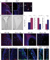
Tlx is required for the generation of adult NSCs. (A) Coronal brain sections of 2-mo-old Tlx+/+ and Tlx−/− mice were stained for GFAP (red). Note that no GFAP+ cells were found in SVZ of Tlx−/− mice, but normal astrocytes can still be found outside the SVZ (arrows). Bar, 30 μm. (B) BrdU. BrdU was administrated to mice for 2 h. Note that no BrdU+ cells in SVZ of Tlx−/− mice are detected. Bar, 30 μm. (C) Number of BrdU+ cells in SVZ per brain section of 2-mo-old Tlx+/+ and Tlx+/− mice. Note that Tlx+/− mice have 38.2 ± 6.7% less labeled cells than Tlx+/+ mice. Mean ± standard deviation (SD). (D) Quantitative PCR validates decreased expression of Tlx mRNA in Tlx+/− (n = 3) versus Tlx+/+ (n = 3) laser-microdissected SVZ cells. Note that expression of Tlx mRNA in Tlx+/− is 48 ± 11% of the wild type. P < 0.01, mean ± standard deviation (SD). (E) Quantitative PCR validates increased expression of PTEN mRNA in Tlx+/− (n = 3) versus Tlx+/+ (n = 3) laser microdissected SVZ cells. Note that expression of PTEN mRNA in Tlx+/− is 78 ± 34% higher than that in Tlx+/+. P < 0.01, mean ± standard deviation (SD). (F, G) Coronal brain sections of P9 Tlx+/+ and Tlx−/− mice were stained for GFAP (red in F). Note that almost no GFAP+ cells were found in the SVZ of Tlx−/− mice. Bar, 10 μm. (G) DCX (red). Note that almost no DCX + cells were found in the SVZ and RMS of Tlx−/− mice. Bar, 30 μm. (H–J) Tamoxifen were injected to Tlx fl/fl; Tlx-CreERT2 and Tlxfl/fl control mice from P5 to P8, the P10 brain sections were stained for GFAP (red in H), Ki67 (green in I), and BLBP (green in J). Note that less GFAP-, Ki67-, and BLBP-expressing cells in the mutant SVZ than the control mice. Bar, 10 μm.

References

    1. Alvarez-Buylla A., Garcia-Verdugo J.M. Neurogenesis in adult subventricular zone. J. Neurosci. 2002;22:629–634. - PMC - PubMed
    1. Anthony T.E., Klein C., Fishell G., Heintz N. Radial glia serve as neuronal progenitors in all regions of the central nervous system. Neuron. 2004;41:881–890. - PubMed
    1. Belz T., Liu H.K., Bock D., Takacs A., Vogt M., Wintermantel T., Brandwein C., Gass P., Greiner E., Schutz G. Inactivation of the gene for the nuclear receptor tailless in the brain preserving its function in the eye. Eur. J. Neurosci. 2007;26:2222–2227. - PubMed
    1. Bickenbach J.R. Identification and behavior of label-retaining cells in oral mucosa and skin. J. Dent. Res. 1981;60:1611–1620. - PubMed
    1. Curtis M.A., Kam M., Nannmark U., Anderson M.F., Axell M.Z., Wikkelso C., Holtas S., van Roon-Mom W.M., Bjork-Eriksson T., Nordborg C., et al. Human neuroblasts migrate to the olfactory bulb via a lateral ventricular extension. Science. 2007;315:1243–1249. - PubMed

Publication types

MeSH terms

Substances