. 2008 Sep 15;22(18):2496-506.
doi: 10.1101/gad.1676108.

PCNA-dependent regulation of p21 ubiquitylation and degradation via the CRL4Cdt2 ubiquitin ligase complex

Affiliations

PCNA-dependent regulation of p21 ubiquitylation and degradation via the CRL4Cdt2 ubiquitin ligase complex

Tarek Abbas et al. Genes Dev. .

Abstract

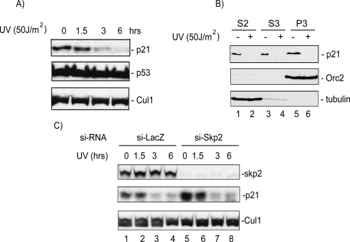
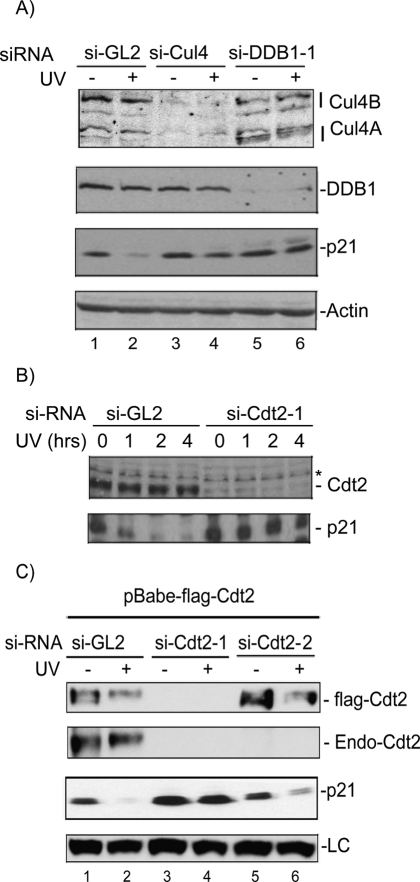
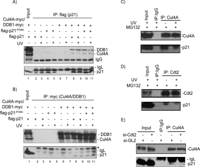
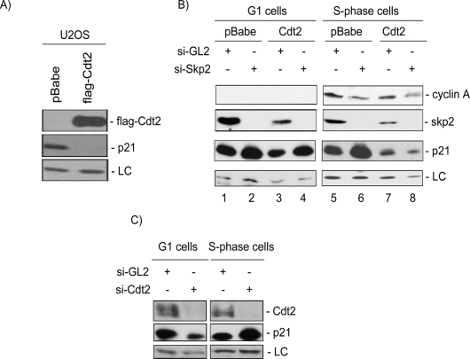
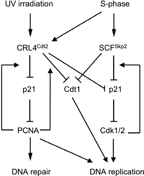
The DNA polymerase delta processivity factor Proliferating Cell Nuclear Antigen (PCNA) promotes the DNA damage-induced degradation of the replication initiation factor Cdt1 via the CRL4(Cdt2) E3 ubiquitin ligase complex. Here we demonstrate that PCNA promotes the ubiquitylation and degradation of the CDK inhibitor p21 in cells irradiated with low dose of ultraviolet (UV) by a similar mechanism. Human cells that are depleted of Cul4, DDB1 (damage-specific DNA-binding protein-1), or the DCAF Cdt2, are deficient in the UV-induced ubiquitylation and degradation of p21. Depletion of mammalian cells of PCNA by siRNA, or mutations in p21 that abrogate PCNA binding, prevent UV-induced p21 ubiquitylation and degradation, indicating that physical binding with PCNA is necessary for the efficient ubiquitylation of p21 via the CRL4(Cdt2) ubiquitin ligase. Cdt2 functions as the substrate recruiting factor for p21 to the rest of the CRL4 ubiquitin ligase complex. The CRL4(Cdt2) E3 ubiquitin ligase ubiquitylates p21 both in vivo and in vitro, and its activity is dependent on the interaction of p21 with PCNA. Finally, we show that the CRL4(Cdt2) and the SCF(Skp2) ubiquitin ligases are redundant with each other in promoting the degradation of p21 during an unperturbed S phase of the cell cycle.


Figures

Figure 1.
Figure 1.
The p21 protein is degraded via a Skp2-independent mechanism in cells irradiated with low-dose UV. (A) Western blot analysis of protein lysate from HeLa cells to test the effect of UV irradiation on p21 degradation, p53 protein level. The CRL1 proteins indicate equal loading. (B) Chromatin-bound p21 is degraded after UV irradiation. Western blot analysis of cytoplasmic (S2), soluble nuclear (S3), and chromatin-bound (P3) lysates of HeLa cells. Anti-Orc2 and anti-tubulin blots demonstrate proper fractionation. (C) A Western blot of HeLa lysates shows that acute silencing of Skp2 by siRNA does not prevent p21 degradation after UV.
Figure 2.
Figure 2.
The CRL4Cdt2 E3 ubiquitin ligase is required to efficiently degrade p21 in UV-irradiated cells. (A) si-RNA knockdown of Cul4A/B or DDB1 prevents p21 degradation after UV. Western blot from nonirradiated or UV-irradiated (20 J/m2) HCT116p53−/− after knockdown of Cul4A and B (si-Cul4), DDB1 (si-DDB1-1), or control siRNA (si-GL2). Actin is shown as a loading control. (B) siRNA knockdown of the DCAF Cdt2 prevents p21 degradation by UV. Western blot of HCT116p53−/− after knockdown of Cdt2 or control si-GL2, UV-irradiated (20 J/m2) and harvested after the indicated hours post-irradiation. (*) A cross-reacting band in the anti-Cdt2 blot that serves as a loading control. (C) Ectopic expression of si-RNA-resistant Cdt2 restores the UV-induced degradation of p21. U2OS cells stably expressing Flag-epitope-tagged Cdt2 (without the 3′UTR) are transfected with either si-GL2 or siRNA targeting the ORF (si-Cdt2-1), or the 3′UTR of Cdt2 (si-Cdt2-2) and UV-irradiated 6 h prior to harvesting. The anti-Cdt2 blot shows endogenous Cdt2 (Endo-Cdt2). An anti-Flag blot shows the exogenously expressed Cdt2 and its efficient silencing by si-Cdt2-1 but not si-Cdt2-2. Exogenous Cdt2 was not shown in the same blot because it overlaps with a cross-reactive band that runs higher than the endogenous protein. (LC) A cross-reacting band in the anti-p21 blot serving as a loading control.
Figure 3.
Figure 3.
p21 interacts with the CRL4Cdt2 ubiquitin ligase in vivo. (A) Ectopically expressed Cul4A-myc and DDB1-myc coimmunoprecipitate with ectopically expressed Flag-p21 or Flag-p21-PCNA. 293T Cells were transiently transfected with plasmids encoding the indicated proteins for 48 h and treated with MG132 for 6 h prior to harvest. Anti-myc Western blot of the anti-Flag immunoprecipitates prepared from 293T cells overexpressing the indicated proteins and shows that Cul4A and DDB1 associate with wild-type p21 and p21-PCNA. An anti-Flag blot demonstrates equal p21 proteins in the anti-Flag immunoprecipitates. (B) The reciprocal of the experiment performed in A, where Flag-p21 (wild type and p21-PCNA) coimmunoprecipitates with CRL4A and DDB1 proteins in the myc IP. (C,D) Endogenous p21 interacts with endogenous Cul4A (C), and Cdt2 (D) in HeLa cells. Lysates of HeLa cells (nonirradiated or UV-irradiated [50 J/m2] and treated with MG132 immediately after irradiation for 6 h prior to harvest) were immunoprecipitated with control anti-IgG (C,D), anti-CRL4A (C), or anti-Cdt2 (D) antibodies. The anti-p21 Western blots show that endogenous p21 is specifically coimmunoprecipitated with Cul4A (C), and Cdt2 (D). (E) Cdt2 mediates the association between p21 and the rest of the CRL4 E3ligase. Proteins extracted from control si-GL2 or si-Cdt2-transfected HeLa cells were immunoprecipitated with the indicated antibodies and immunoblotted with anti-Cul4 or anti-p21. The results show that endogenous p21 protein associates with endogenous Cul4A only in the presence of Cdt2.
Figure 4.
Figure 4.
PCNA promotes the polyubiquitylation and degradation of p21 after UV irradiation, and its physical interaction with p21 is necessary for the degradation. (A) si-RNA knockdown of PCNA prevents p21 degradation in UV-irradiated cells. Western blot of lysates of HCT116p53−/− cells transfected with si-GL2 control oligos or siRNA targeting PCNA (si-PCNA) and immunoblotted for the indicated proteins. (B) p21-PCNA does not interact with PCNA. Cell lysates and Flag immunoprecipitates Western blotted for Flag-p21 and PCNA. (C) Western blot of HeLa cells transfected with Flag-p21 or Flag-p21-PCNA with or without exposure to UV. (LC) A cross-reacting band serving as a loading control. (D) Time-course analysis of the degradation of ectopically expressed p21 and p21-PCNA proteins in HeLa cells following UV (20 J/m2). The anti-p21 blot indicates that endogenous wild-type p21 is visibly degraded within 2 h of UV irradiation. Flag-p21-PCNA remains stable for up to 8 h following UV. (E) Quantitation of Flag-p21 proteins displayed in D to determine the rate of degradation of p21 relative to p21-PCNA in response to UV irradiation. (F) Wild-type p21 but not p21-PCNA is efficiently degraded at various low doses of UV irradiation. HCT116p21−/− stably expressing either Flag-p21 or Flag-p21-PCNA harvested 4 h after radiation and immunoblotted for Flag. (*) A cross-reacting band serving as a loading control. (G) Wild-type p21 but not p21-PCNA is efficiently ubiquitylated in vivo and that the ubiquitylation is stimulated upon UV irradiation. (Top) Anti-p21 Western blot analysis of the immunoprecipitated ubiquitylated proteins (with anti-HA antibody) extracted from HCT116p21−/− coexpressing wild-type p21 or p21-PCNA and HA-tagged ubiquitin. (Bottom) Anti-p21 Western blot of the anti-Flag immunoprecipitates demonstrates that p21 and p21-PCNA were expressed to similar levels in these cells. A low exposure of the nonubiquitylated p21 is shown as well. The X-ray films were scanned, and the ratio of the ubiquitylated p21 to that of total p21 (ubiquitylated + nonubiquitylated p21 from the nonsaturating lower-exposure film) is indicated below each lane.
Figure 5.
Figure 5.
CRL4Cdt2 E3 ubiquitin ligase promotes p21 ubiquitylation in vitro. (A,B) The CRL4Cdt2 E3 ubiquitin ligase complex polyubiquitylates p21 in vitro. CRL4Cdt2 enzyme complexes (with the indicated overexpressed proteins on top) were immuno-purified using either (A) anti-Flag or (B) anti-myc antibodies and mixed with separately immuno-purified p21 substrate in an in vitro ubiquitylation reaction. Western blots for DDB1 and CRL4A (anti-myc), Cdt2 (anti-Cdt2) demonstrate the formation of the stable E3 ligase in 293T cells used for the enzyme complex preparation. The anti-PCNA blot shows that PCNA copurified with p21. Anti-p21 immunoblots show the formation of higher p21 molecular weight species corresponding to polyubiquitylated p21. (C) Cdt2 containing E3 ligase is capable of ubiquitylating p21 in vitro. Flag-Cdt2 expressed stably in 293T cells, immunoprecipitated with anti-Flag agarose and incubated with separately purified p21 protein in an in vitro ubiquitylation reaction. Mock IP from control 293T cells were used as a negative control. A high exposure of the anti-p21 blot is shown (right) with ubiquitylated p21 labeled with asterisks (*). The results demonstrate that CRL4Cdt2 is sufficient to polyubiquitylate p21 in vitro.
Figure 6.
Figure 6.
PCNA is required for the efficient ubiquitylation of p21 in vitro and p21 ubiquitylation is enhanced by phosphomimetic substitution of p21 at Ser114. (A) In vitro ubiquitylation of p21 via the CRL4Cdt2 E3 ligase purified from cells that were depleted of PCNA by siRNA and overexpressing Cul4A, DDB1, Cdt2, and Rbx1 (CRL4Cdt2). The p21 substrate was purified separately from cells also depleted of PCNA. The Western blot demonstrates that p21 is ubiquitylated via the CRL4Cdt2 E3 only when the substrate, p21, copurified with PCNA. The anti-PCNA Western blot shows that PCNA was absent in the substrate preparation from 293T cells that were treated with si-PCNA. (B) Wild-type p21 but not p21-PCNA is ubiquitylated via the CRL4Cdt2 E3 ligase. Time-course analysis of the CRL4Cdt2 E3-mediated ubiquitylation of p21 proteins in vitro. The anti-p21 blot shows that p21 and p21-PCNA are equal in both reaction sets. Time indicates the incubation time in minutes. The anti-PCNA blot shows that PCNA copurifies only with wild type but not the p21-PCNA mutant. (C) In vitro ubiquitylation of wild-type p21 (lanes 1–3) or p21 with a phosphomimetic substitution at Ser114 (p21S114E) (lane 4) via the CRL4Cdt2 ubiquitin ligase. (Lane 5) In vitro ubiquitylation of wild-type p21 via immuno-purified CRL1 E3 (Skp2-dependent ligase) is also shown for comparison. The Western blot was probed for p21 to demonstrate the formation of p21 polyubiquitin chains. The p21S114E runs slightly higher than wild-type protein. A low exposure of the anti-p21 blot demonstrates equal p21 and p21S114E substrates.
Figure 7.
Figure 7.
Cdt2 promotes the destruction of p21 in S-phase cells. (A) Stable expression of Cdt2 significantly reduces the steady-state levels of p21 in asynchronous U2OS cells. Soluble protein lysates of Mock or Cdt2-overexpressing U2OS cells were resolved on SDS-PAGE and analyzed for Cdt2 (anti-Flag) and p21 protein levels. (LC) A cross-reacting band in the anti-p21 blot serving as a loading control. (B) Cdt2 specifically targets p21 in S phase of the cell cycle. Control or Cdt2 overexpressing U2OS cells were transfected either with control siRNA (si-GL2) or with siRNA against Skp2. Forty-eight hours after transfections, cells were synchronized with nocodazole, and mitotic cells were collected by shake-off, washed with PBS, and incubated for an additional 8 h (G1 cells) or 16 h (S-phase cells), and the cell lysates were analyzed for the indicated proteins. Cyclin A levels show the synchronization of cells in G1 (left) and S phase (right) of the cell cycle. The results demonstrate that Skp2 down-regulation by siRNA results in the accumulation of p21 in both G1- and S-phase cells. On the other hand, ectopic expression of Cdt2 significantly reduced p21 only in S-phase cells, and that reduction occurred even after down-regulating Skp2 (lanes 7,8). (C) Endogenous Cdt2 promotes the destruction of p21 during S phase of the cell cycle. HeLa cells transfected with either control si-RNA (si-GL2) or siRNA targeting Cdt2 (si-Cdt2) and synchronized in G1 and S phases of the cell cycle as described in B. Protein lysates were subsequently analyzed as in B. The anti-Cdt2 blot demonstrates efficient down-regulation of Cdt2 in cells transfected with si-Cdt2. (LC) A cross-reactive band in the anti-p21 blot used as loading control. The results demonstrate that p21 protein is specifically stabilized in S-phase cells but not in cells in the G1 phase of the cell cycle upon depletion of Cdt2.
Figure 8.
Figure 8.
Model of the dual role for the CRL4Cdt2 E3 ubiquitin ligase in response to low-dose UV irradiation and during the S phase of the cell cycle. The E3 ubiquitin ligase targets the replication factor Cdt1 for degradation, thereby suppressing replication licensing until the damage is repaired or until the G1 phase of the next cell cycle. Meanwhile, the same E3 ubiquitin ligase targets the Cdk inhibitor p21 for degradation, thereby allowing PCNA to function free of inhibitory p21 in promoting DNA repair or to allow the cells to progress through the S phase in the absence of DNA damage (the latter function being partially redundant with the Skp2-mediated degradation of p21). Both activities are dependent on the physical interaction of both of these substrates to PCNA.

References

    1. Amador V., Ge S., Santamaria P.G., Guardavaccaro D., Pagano M. APC/C(Cdc20) controls the ubiquitin-mediated degradation of p21 in prometaphase. Mol. Cell. 2007;27:462–473. - PMC - PubMed
    1. Arias E.E., Walter J.C. PCNA functions as a molecular platform to trigger Cdt1 destruction and prevent re-replication. Nat. Cell Biol. 2006;8:84–90. - PubMed
    1. Banks D., Wu M., Higa L.A., Gavrilova N., Quan J., Ye T., Kobayashi R., Sun H., Zhang H. L2DTL/CDT2 and PCNA interact with p53 and regulate p53 polyubiquitination and protein stability through MDM2 and CUL4A/DDB1 complexes. Cell Cycle. 2006;5:1719–1729. - PubMed
    1. Bendjennat M., Boulaire J., Jascur T., Brickner H., Barbier V., Sarasin A., Fotedar A., Fotedar R. UV irradiation triggers ubiquitin-dependent degradation of p21(WAF1) to promote DNA repair. Cell. 2003;114:599–610. - PubMed
    1. Bornstein G., Bloom J., Sitry-Shevah D., Nakayama K., Pagano M., Hershko A. Role of the SCFSkp2 ubiquitin ligase in the degradation of p21Cip1 in S phase. J. Biol. Chem. 2003;278:25752–25757. - PubMed

Publication types

Substances