. 2008 Nov;28(22):6773-84.
doi: 10.1128/MCB.00941-08. Epub 2008 Sep 15.

MicroRNA-155 is regulated by the transforming growth factor beta/Smad pathway and contributes to epithelial cell plasticity by targeting RhoA

Affiliations

MicroRNA-155 is regulated by the transforming growth factor beta/Smad pathway and contributes to epithelial cell plasticity by targeting RhoA

William Kong et al. Mol Cell Biol. 2008 Nov.

Abstract

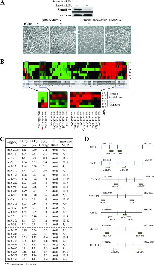
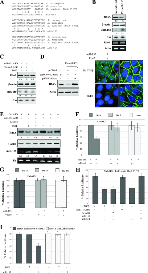
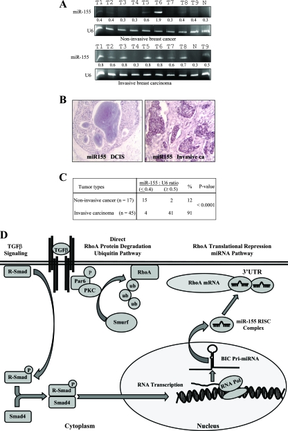
Transforming growth factor beta (TGF-beta) signaling facilitates metastasis in advanced malignancy. While a number of protein-encoding genes are known to be involved in this process, information on the role of microRNAs (miRNAs) in TGF-beta-induced cell migration and invasion is still limited. By hybridizing a 515-miRNA oligonucleotide-based microarray library, a total of 28 miRNAs were found to be significantly deregulated in TGF-beta-treated normal murine mammary gland (NMuMG) epithelial cells but not Smad4 knockdown NMuMG cells. Among upregulated miRNAs, miR-155 was the most significantly elevated miRNA. TGF-beta induces miR-155 expression and promoter activity through Smad4. The knockdown of miR-155 suppressed TGF-beta-induced epithelial-mesenchymal transition (EMT) and tight junction dissolution, as well as cell migration and invasion. Further, the ectopic expression of miR-155 reduced RhoA protein and disrupted tight junction formation. Reintroducing RhoA cDNA without the 3' untranslated region largely reversed the phenotype induced by miR-155 and TGF-beta. In addition, elevated levels of miR-155 were frequently detected in invasive breast cancer tissues. These data suggest that miR-155 may play an important role in TGF-beta-induced EMT and cell migration and invasion by targeting RhoA and indicate that it is a potential therapeutic target for breast cancer intervention.


Figures

FIG. 1.
FIG. 1.
Profile of miRNA expression in TGF-β/Smad-induced EMT in NMuMG cells. (A) TGF-β induces the cellular morphological change of EMT in control (pRS vector-transfected) but not Smad4 knockdown NMuMG cells. (Upper panels) A Western blot analysis of control and Smad4-knockdown NMuMG cells was performed with anti-Smad4 and antiactin antibodies. shRNA, short hairpin RNA; +, present; −, absent. (Lower panels) The indicated cells were treated with (+) or without (−) TGF-β for 24 h and photographed. pRS-NMuMG, pRS vector-transfected NMuMG cells. (B) Heat map representation of miRNAs deregulated in control and Smad4 knockdown NMuMG cells during TGF-β treatment. The red arrowhead indicates hsa-miR-155 highest upregulated miRNA. (C) List of deregulated miRNAs induced by TGF-β in control but not Smad4 knockdown NMuMG cells. (D) Chromosomal representation of the locations of deregulated miRNAs within mouse genomic DNA. Clustered miRNAs were simultaneously downregulated or upregulated during TGF-β treatment. Chr, chromosome.
FIG. 2.
FIG. 2.
TGF-β/Smad transcriptionally regulates miR-155. (A) Verification of TGF-β-regulated miRNAs. Parental NMuMG cells were treated with TGF-β for the indicated times and subjected to Northern blot analysis with the indicated probes. The numbers between the gels represent the miR-155-to-U6 band density ratios. (B) TGF-β induces miR-155 promoter activity in parental but not Smad4 knockdown NMuMG cells. (Top panel) The diagram depicts the putative mouse miR-155 promoter construct containing two Smad4-binding sites and the individual Smad4-binding-site deletion mutant constructs cloned into the pGL3 plasmid. Parental and Smad4 knockdown cells were transfected with pGL3-miR-155-Luc and treated with or without TGF-β. Following 36 h of incubation, the cells were subjected to a luciferase reporter assay. The experiments were done three times with triplicate samples for each treatment. +, present; −, absent. (C) The first Smad4-binding site is required for TGF-β-induced miR-155 promoter activity. NMuMG cells were transfected with the indicated plasmids, treated (+) with TGF-β or left untreated (−), and assayed for luciferase activity. Values are expressed as relative luciferase units. (D) TGF-β induces Smad4 binding to the miR-155 promoter. NMuMG cells treated with or without TGF-β were evaluated by a ChIP assay. PCR was done with the eluted DNA fragments from anti-Smad4 immunoprecipitates by using a set of primers that detect the first Smad4-binding site, determined in the reporter assay to be important. IgG and antiactin antibody were used as negative controls. α-Smad4, anti-Smad4 antibody.
FIG. 3.
FIG. 3.
miR-155 mediates the effect of TGF-β on EMT. (A) Ectopic expression and knockdown of miR-155. NMuMG cells were transfected with the indicated plasmids and oligonucleotides. Following treatment with (+) or without (−) TGF-β, cells were subjected to Northern blot analysis with [α-32P]dATP-labeled miR-155 and U6 probes. Pre miR-155, pcDNA6.2-GW/miR-155; Pre miR-ctrl, pcDNA6.2-GW/miR-control. (B and C) The knockdown of miR-155 inhibited TGF-β-induced EMT and tight junction dissolution. NMuMG cells were transfected with miR-155 ASO and control ASO and then treated with or without TGFβ for 24 h. Cell morphologies were documented using a phase-contrast microscope (B), and cells were stained with anti-ZO-1 and anti-E-cadherin antibodies conjugated to fluorescein isothiocyanate and tetramethyl rhodamine isothiocyanate, respectively (C). Arrows in panel C indicate the restoration of TGF-β-disrupted tight junctions by the knockdown of miR-155. (D) The knockdown of miR-155 inhibits the TGF-β downregulation of E-cadherin (E-Cad). NMuMG cells were transfected with control and miR-155 ASO, treated with TGF-β for 24 h, and then immunoblotted with the indicated antibodies. (E and F) The overexpression of miR-155 disrupted proper tight junction formations and accelerated TGF-β-induced EMT. Stably miR-155-transfected and control NMuMG cells were treated with or without TGF-β for 12 h and photographed (E) and immunofluorescence-stained with the indicated antibodies (F). The tight junction dissolution promoted by the ectopic expression of miR-155 is indicated by arrows. (G) Western blot analysis of E-cadherin in NMuMG cells that were transfected with miR-155 and then treated with TGF-β for 12 h.
FIG. 4.
FIG. 4.
miR-155 plays a significant role in cell migration and invasion. (A and B) The ectopic expression of miR-155 induces cell migration and invasion. NMuMG cells stably transfected with pcDNA6.2-GW/miR-control (Pre miR-ctrl) and pcDNA6.2-GW/miR-155 (Pre miR-155) were examined for cell migration and invasion by using Boyden chamber and wound healing assays as previously described (28). (C) The knockdown of miR-155 reduces cell migration and invasion. NMuMG cells were transfected with miR-155 ASO and control ASO. Following 36 h of incubation, cells were subjected to chamber cell migration and invasion assays using Boyden chambers with or without the inclusion of coating Matrigel. (D) Statistical analysis. The experiments depicted in panels A to C were repeated three times. P values for comparisons are indicated. Values are arbitrary units.
FIG. 5.
FIG. 5.
RhoA is a target of miR-155. (A) Sequence alignment of miR-155 with the 3′ UTR of the RhoA gene. The seed sequence of miR-155 matches three regions of the RhoA gene 3′ UTR, which are highly conserved among humans (Homo sapiens), mice (Mus musculus), and rats (Rattus norvegicus). Vertical lines and colons indicate Watson-Crick and wobble base pairing, respectively. (B) miR-155 reduces RhoA protein but not mRNA expression. NMuMG cells were transfected with pcDNA6.2-GW/miR-155 (Pre miR-155) and control vector (Pre miR-Ctrl). After selection with blasticidin, the expression level of RhoA was determined using Western blot analysis (first panel). The same blot was reprobed with antiactin antibody (second panel). The expression of miR-155 from the same set of cells was examined by Northern blot analysis (third panel). RT-PCR was performed to determine RhoA mRNA levels (fifth panel). U6 and actin were used for loading controls (fourth and sixth panels). The numbers between the gels represent the miR-155-to-U6 band density ratios. (C) The knockdown of miR-155 inhibits the TGF-β downregulation of RhoA. NMuMG cells were transfected with miR-155 ASO or control ASO. After 36 h of incubation, cells were treated with (+) or without (−) TGF-β for 24 h and immunoblotted with the indicated antibodies (first and second panels). A Northern blot was hybridized with the indicated probes (third and fourth panels). (D) The ectopic expression of RhoA cDNA lacking the 3′ UTR overrides the effects of miR-155 on cell tight junction dissolution. Clonal cells stably expressing miR-155 were transfected with pcDNA3-RhoA or control vector. Following treatment with or without TGF-β, cells were immunostained with anti-ZO-1 antibody. (E) The inhibition of the ubiquitin-proteasome pathway and miR-155 hinders TGF-β downregulation of RhoA. NMuMG cells were transfected with control (ctrl) or miR-155 ASO. Following 48 h of incubation, cells were treated with or without MG132 and/or TGF-β for 12 h and then subjected to immunoblotting (first and second panels) and qRT-PCR (third and fourth panels). (F and G) miR-155 inhibits RhoA gene 3′ UTR luciferase activity. Cells were transfected with individual site constructs in pGL3 (F) and the corresponding mutant constructs (G), together with pCMV-β-galactosidase, pcDNA6.2-GW/miR-155 (miR-155), or pcDNA6.2-GW/miR-control vector (miR-ctrl). Luciferase activities were normalized to β-galactosidase activity. (H and I) TGF-β and miR-155 repress the full-length RhoA gene 3′ UTR but not its antisense strand in parental NMuMG cells. Cells were transfected with the indicated plasmids and treated with or without TGF-β for 24 h. Luciferase activities were normalized to β-galactosidase activity. The full-length RhoA gene 3′ UTR responds to TGF-β and miR-155 in parental (H) but not Smad4 knockdown (I) NMuMG cells. TGF-β-inhibited reporter activity was inhibited by the knockdown of miR-155. miR-214 was used as a control. Experiments were done in triplicate for standard deviation calculations. RhoA 3′ UTR-AS/NMuMG, NMuMG cells transfected with the antisense strand of the RhoA gene 3′ UTR.
FIG. 6.
FIG. 6.
Elevated levels of miR-155 are associated with invasive breast cancer. (A) qRT-PCR analysis of miR-155 expression in normal human breast tissue (lanes N), noninvasive breast cancer tissue, and invasive breast carcinoma. U6 was used as a control. The numbers between the gels represent the miR-155-to-U6 band density ratios. T1 to T9 indicate the tumor samples. (B) LNA-ISH analyses. miR-155 was labeled with digoxigenin-ddUTP by using the Dig 3′-end labeling kit (Roche) and hybridized to paraffin sections of breast cancer tissue. Representative photomicrographs of sections of ductal carcinoma in situ (DCIS) breast tissue (left) and invasive breast tumor tissue (right) are shown. (C) Summary of qRT-PCR and LNA-ISH analyses. miR-155 was detected more frequently in invasive breast carcinoma tissue than in noninvasive tumor tissue. (D) Schematic illustration of the transcriptional induction of miR-155 by the TGF-β/Smad pathway and the TGF-β downregulation of RhoA through the ubiquitin-proteasome and miR-155 cascades. PKC, protein kinase C; RISC, RNA-induced silencing complex; RNA Pol, RNA polymerase.

References

    1. Ambros, V. 2004. The functions of animal microRNAs. Nature 431350-355. - PubMed
    1. Burk, U., J. Schubert, U. Wellner, O. Schmalhofer, E. Vincan, S. Spaderna, and T. Brabletz. 2008. A reciprocal repression between ZEB1 and members of the miR-200 family promotes EMT and invasion in cancer cells. EMBO Rep. 9582-589. - PMC - PubMed
    1. Cai, X., C. H. Hagedorn, and B. R. Cullen. 2004. Human microRNAs are processed from capped, polyadenylated transcripts that can also function as mRNAs. RNA 101957-1966. - PMC - PubMed
    1. Costinean, S., N. Zanesi, Y. Pekarsky, E. Tili, S. Volinia, N. Heerema, and C. M. Croce. 2006. Pre-B cell proliferation and lymphoblastic leukemia/high-grade lymphoma in Eμ-miR155 transgenic mice. Proc. Natl. Acad. Sci. USA 1037024-7029. - PMC - PubMed
    1. Deckers, M., M. van Dinther, J. Buijs, I. Que, C. Lowik, G. van der Pluijm, and P. ten Dijke. 2006. The tumor suppressor Smad4 is required for transforming growth factor beta-induced epithelial to mesenchymal transition and bone metastasis of breast cancer cells. Cancer Res. 662202-2209. - PubMed

Publication types

MeSH terms

LinkOut - more resources