. 2008 Nov;28(22):6954-66.
doi: 10.1128/MCB.00925-08. Epub 2008 Sep 15.

Extracellular signal-regulated kinase 2 (ERK2) phosphorylation sites and docking domain on the nuclear pore complex protein Tpr cooperatively regulate ERK2-Tpr interaction

Affiliations

Extracellular signal-regulated kinase 2 (ERK2) phosphorylation sites and docking domain on the nuclear pore complex protein Tpr cooperatively regulate ERK2-Tpr interaction

Tomás Vomastek et al. Mol Cell Biol. 2008 Nov.

Abstract

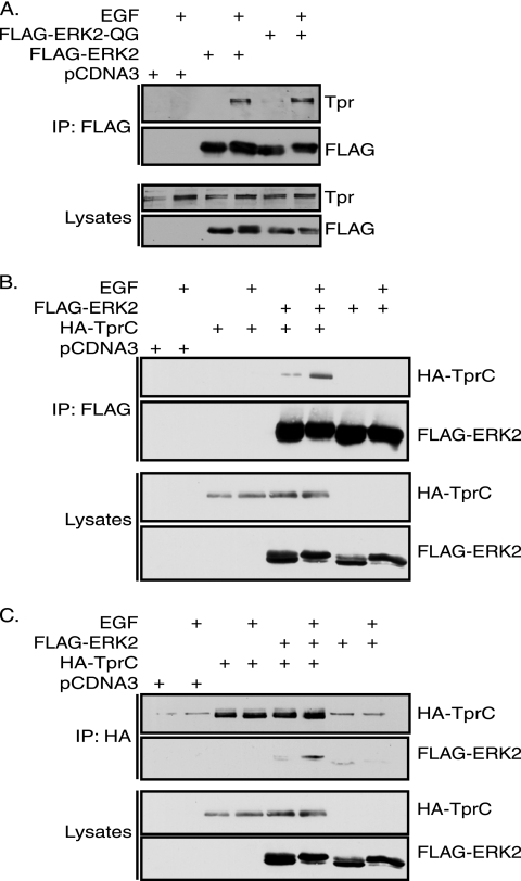
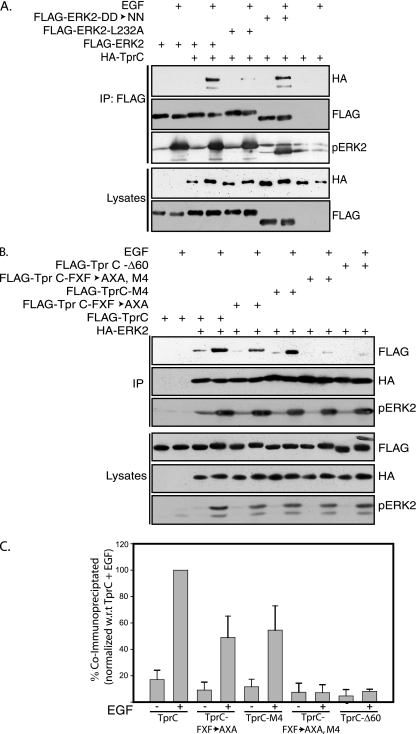
Identifying direct substrates of mitogen-activated protein kinases (MAPKs) and understanding how those substrates are selected is central to understanding how these ubiquitously activated enzymes generate diverse biological responses. In previous work, we identified several new candidate substrates for the MAPK ERK2 (extracellular signal-regulated kinase 2), including the nuclear pore complex protein Tpr (translocated promoter region). In this report, we identify sites on Tpr for ERK2 phosphorylation and binding and demonstrate their functional interaction. ERK2 phosphorylation and dimerization are necessary for ERK2-Tpr binding, and this occurs through a DEF (docking site for ERK2, FXF) domain on Tpr. Surprisingly, the DEF domain and the phosphorylation sites displayed positive cooperativity to promote ERK2 binding to Tpr, in contrast to substrates where phosphorylation reduces binding. Ectopic expression or depletion of Tpr resulted in decreased movement of activated ERK2 from the cytoplasm to the nucleus, implying a role for Tpr in ERK2 translocation. Collectively, the data provide direct evidence that a component of the nuclear pore complex is a bona fide substrate of ERK2 in vivo and that activated ERK2 stably associates with this substrate after phosphorylation, where it could play a continuing role in nuclear pore function. We propose that Tpr is both a substrate and a scaffold for activated ERKs.


Figures

FIG. 1.
FIG. 1.
Carboxy terminus of Tpr is sufficient for association with ERK2 upon EGF stimulation. (A) Cells transfected with pcDNA3, FLAG-ERK2, and FLAG-ERK2-QG were serum starved for 4 to 5 h followed by stimulation with EGF (20 ng/ml) for 10 min. Cells were lysed and proteins immunoprecipitated (IP) with FLAG-M2 agarose beads. Immunoprecipitated proteins were resolved by 10% SDS-PAGE, followed by immunoblotting with anti-FLAG-M2 or anti-Tpr antibodies. (B) Cells transfected with pcDNA3, HA-TprC, FLAG-ERK2, or FLAG-ERK2 and HA-TprC together were serum starved and stimulated with EGF. Proteins were immunoprecipitated and analyzed by immunoblotting as described for panel A and probed with anti-FLAG-M2 and anti-HA antibodies. (C) As in panel B, except that the proteins were immunoprecipitated with anti-HA antibodies coupled to protein A agarose, followed by immunoblotting with anti-FLAG-M2 and anti-HA antibodies.
FIG. 2.
FIG. 2.
Tpr does not interact with MEK1, JNK1, or p38α. (A) Cells transfected with pcDNA3, FLAG-ERK2, FLAG-ERK2-QG, FLAG-ERK1, and FLAG-MEK1 were serum starved and stimulated with EGF. Cells were lysed and proteins immunoprecipitated (IP) with FLAG-M2 agarose beads followed by immunoblotting with anti-Tpr and anti-FLAG-M2 antibodies. (B) Cells were transfected with pcDNA3 or HA-TprC, or double transfected with HA-TprC and FLAG-ERK2 or FLAG-JNK1 or FLAG-p38α. The last two were stimulated with anisomycin for 30 min instead of EGF. Cell lysates were probed with anti-FLAG-M2 and anti-HA antibodies. Based on these results, twice the amount of lysate was used for immunoprecipitation from cells cotransfected with FLAG-JNK1 and HA-TprC, compared to amounts for other transfected cells. Proteins were immunoprecipitated with FLAG-M2 agarose beads. One-twentieth of the immunoprecipitated proteins was analyzed by probing with anti-FLAG-M2, anti-phosphor-ERK2 (Sigma), anti-phosphor-JNK1 (Sigma), and anti-phospho-p38α (Sigma). The remaining immunoprecipitated proteins were probed with anti-HA antibodies.
FIG. 3.
FIG. 3.
ERK2 phosphorylates serine and threonine residues of Tpr in vitro. (A) Cells were transfected individually with FLAG-ERK2, FLAG-TprC, and pcDNA3 vectors. Cells were serum starved, and FLAG-ERK2-transfected cells were stimulated with EGF. Proteins were immunoprecipitated from lysed cells with FLAG-M2 agarose beads, and FLAG-ERK2 immunoprecipitate was mixed either with FLAG-TprC immunoprecipitate or with immunoprecipitate from pcDNA3-transfected cells. Kinase reactions were carried out with [γ-32P]ATP at 30°C for 10 min. (B) Phosphoamino acid analysis of in vitro-labeled TprC from time course reactions. p, phosphorylated. (C) In vitro phosphorylated TprC was digested with trypsin, and the resulting phosphopeptides were mapped by two-dimensional TLC (4). Two major spots are indicated by white arrows, and four minor spots are indicated by black arrows.
FIG. 4.
FIG. 4.
Tpr is phosphorylated by ERK2 at four different sites. (A) COS-1 cells were transfected with FLAG-TprFL, FLAG-TprC, and FLAG-TprC mutants (as specified above the maps). The immunoprecipitated FLAG-tagged Tpr proteins were mixed with immunoprecipitated FLAG-ERK2, and kinase reactions were carried out with [γ-32P]ATP. In vitro-phosphorylated FLAG-tagged TprFL, TprC, and TprC mutants were digested with trypsin, and the resulting phosphopeptides were mapped by two-dimensional separation on TLC. Spots in similar positions in TprFL and TprC maps are indicated by numbered black arrows. Spots missing due to mutations in the TprC phosphorylation sites are indicated by numbered white arrows. (B) The upper panel depicts the nomenclature of the combination target site mutants of TprC. The putative D domain (2091RRQSVGRGLQL2101) and DEF motif (2150FRF2152) are also indicated (residues that match the consensus for each motif are indicated by bold). In vitro-phosphorylated proteins were digested with trypsin, and the resulting phosphopeptides were mapped by TLC (4). Major (spots 3, 4, and 4*) and minor (spots 1 and 2) spots in TprC are indicated by numbered black arrows. Missing spots in TprCM2, TprC16M3, and TprC12M3 are indicated by numbered white arrows. In TprC-M4, the location of the missing spots is circled by a dotted line. (C) Incorporation of 32P in in vitro-phosphorylated TprC, TprC-M4, and the vector control samples was quantified by Cerenkov counting in three independent experiments. Counts in TprC were normalized to 100% in each experiment, and the percent counts in other samples were calculated with respect to TprC. The results were plotted with percent counts with respect to TprC on the y axis and the samples on the x axis.
FIG. 5.
FIG. 5.
ERK2 phosphorylates Tpr at the same sites in vivo and in vitro. (A) COS-1 cells were transfected with FLAG-TprFL or FLAG-TprC vectors. Transfected cells were labeled metabolically for 3 h. For the EGF-stimulated samples, EGF (20 ng/ml) was added after 3 h to the labeling cells for 10 min. For samples with MEK inhibitor UO126, 20 μM of inhibitor was present during the 3-h labeling and EGF stimulation. FLAG-tagged TprFL and TprC were immunoprecipitated, resolved, transferred, and autoradiographed. In vivo-labeled TprFL and TprC were digested with trypsin, and the resulting phosphopeptides were mapped by two-dimensional TLC. In vitro-labeled TprC from the earlier experiment was resolved on TLC plates along with in vivo-labeled TprC and TprFL. Peptide spots that show slight increases in intensity following EGF stimulation are indicated by white arrows (panel 2). Spots that did not disappear with UO126 pretreatment are marked in the first panel by a dotted line. (B) FLAG-TprC- and FLAG-TprC-M4-transfected cells were metabolically labeled as described above with EGF stimulation. Tryptic digested peptides were resolved by two-dimensional TLC (4).
FIG. 6.
FIG. 6.
Tpr-ERK2 interaction requires ERK2 phosphorylation and dimerization, but not the D domain. (A) Cells transfected with pcDNA3, FLAG-ERK2, and the FLAG-ERK2-TAYF mutant were serum starved and stimulated with EGF. Proteins immunoprecipitated (IP) with FLAG-M2 agarose beads from lysed cells were immunoblotted and probed with anti-FLAG and anti-Tpr antibodies. (B) Cells transfected with pcDNA3, FLAG-ERK2, and the FLAG-ERK2-Δ4 dimerization mutant were serum starved and stimulated with EGF. Proteins immunoprecipitated with FLAG-M2 agarose beads were immunoblotted and probed with anti-FLAG and anti-Tpr antibodies. (C) COS-1 cells transfected with pcDNA3, FLAG-ERK2, and FLAG-ERK2-DD→NN were serum starved, followed by stimulation with EGF as usual. Proteins immunoprecipitated from lysed cells with α-FLAG-M2 agarose beads were immunoblotted, and lysates and immunoprecipitates were probed with anti-FLAG and anti-MEK1 mouse monoclonal (Transduction Laboratories), anti-Rsk1 goat polyclonal (Santa Cruz Biotechnology Inc.), and anti-Tpr antibodies.
FIG. 7.
FIG. 7.
ERK2 interacts with Tpr through DEF and phosphorylation sites. (A) Cells were cotransfected with FLAG-ERK2 or FLAG-ERK2-DD→NN or FLAG-ERK2 L232A or pcDNA3 and HA-TprC. Following serum starvation and EGF stimulation, proteins were immunoprecipitated (IP) with anti-FLAG antibodies. Lysates and immunoprecipitated proteins were probed with anti-FLAG and anti-HA antibodies. Immunoprecipitates were also probed with α-phospho-ERK antibodies. (B) Cells were transfected with FLAG-TprC or HA-ERK2 or double transfected with FLAG-TprC or FLAG-TprC-FXF→AXA, FLAG-TprC-M4, or FLAG-TprC-FXF→AXA,M4 or FLAG-TprC-Δ60 and HA-ERK2. Following serum starvation and EGF stimulation, proteins were immunoprecipitated with anti-HA antibodies. Lysates and immunoprecipitated proteins were probed with anti-FLAG, anti-HA, and anti-phospho-ERK antibodies. (C) Appropriate enhanced chemiluminescence exposures that are within the linear range of the X-ray film were used for quantitating coimmunoprecipitated wild-type and Tpr mutants using Image J software (Wayne Rasband, National Institutes of Health). Presence (+) or absence (−) of EGF and the sample names are indicated. Values obtained for the TprC+EGF sample were set at 100%. Percent binding values for the remaining samples were calculated with respect to TprC+EGF in each experiment. Percent binding values from three independent experiments were used for calculating standard deviations.
FIG. 8.
FIG. 8.
Tpr phosphorylation enhances ERK2-Tpr binding. (A) Cells were cotransfected with pcDNA3 or HA-ERK2 or HA-ERK2-K52R and FLAG-TprC or FLAG-TprC-FXF→AXA. Following serum starvation and EGF stimulation, proteins were immunoprecipitated (IP) with anti-HA beads (Sigma). Lysates and immunoprecipitated proteins were probed with anti-FLAG and anti-HA antibodies.
FIG. 9.
FIG. 9.
ERK2 phosphorylates proteins that interact with Tpr. (A) COS-1 cells were cotransfected with FLAG-TprFL or FLAG-TprFL-FXF→AXA,M4 and HA-ERK2 or HA-ERK2-QG. Cells were serum starved and stimulated with 20 ng/ml EGF, and the proteins were immunoprecipitated (IP) with FLAG-M2 agarose beads. In vitro kinase reactions were carried out with 1/10 of the immunoprecipitated proteins, and part of the lysate was resolved by SDS-PAGE, transferred onto a nitrocellulose membrane, and probed with anti-FLAG and anti-HA antibodies. (B) Kinase reactions were carried out with the remaining 9/10 of immunoprecipitated proteins by using ∼20 μCi [γ-32P]cpATP (analog ATP) as the substrate for 15 min at 30°C. One-fifth of the volume of these reaction mixtures was resolved by SDS-PAGE, transferred onto a nitrocellulose membrane, and autoradiographed. (C) The remaining part of the kinase reaction mixtures was resolved on pH 4 to 7 two-dimensional gels as described in our previous report (10). Background spots indicated by white arrows were used for aligning the autoradiograms. Additional spots that are specifically detected in TprFL+ERK2-QG reactions (middle panel) are indicated by black arrows.
FIG. 10.
FIG. 10.
Tpr regulates YFP-ERK recruitment into the nucleus in response to EGF stimulation. Control- or Tpr siRNA-treated HEK293T cells expressing CFP-MEK and YFP-ERK were plated on glass dishes coated with extracellular matrix components (10 μg/ml fibronectin and 5 μg/ml collagen type IV). The next day, cells were serum deprived for 3 h and stimulated with EGF (10 ng/ml). (A) Western blot analysis of Tpr expression in control and Tpr siRNA-treated cells. Lysates from control and Tpr knockdown cells were probed with antiTpr and antipaxillin antibody as described in Materials and Methods. (B) Images of and quantification of YFP-ERK nuclear localization in control and Tpr siRNA-treated HEK293T cells prior to and after EGF stimulation. (C) Images show the quantification of YFP-ERK in pcDNA3- or Flag-Tpr-expressing cells prior to and after EGF stimulation and Western blot analysis of Flag-Tpr expression in pcDNA3- and Flag-Tpr-expressing cells. In panels B and C, approximately 50 to 70 cells per each experimental condition were used to quantify YFP-ERK translocation to the nucleus. Each experiment was repeated twice.
FIG. 11.
FIG. 11.
Localization of GFP-Tpr and GFP-Tpr-M4. Images show the confocal microscopy of COS-1 cells transfected with GFP-Tpr or GFP-Tpr-M4 (as described in Materials and Methods).

References

    1. Adachi, M., M. Fukuda, and E. Nishida. 1999. Two co-existing mechanisms for nuclear import of MAP kinase: passive diffusion of a monomer and active transport of a dimer. EMBO J. 185347-5358. - PMC - PubMed
    1. Bangs, P., B. Burke, C. Powers, R. Craig, A. Purohit, and S. Doxsey. 1998. Functional analysis of Tpr: identification of nuclear pore complex association and nuclear localization domains and a role in mRNA export. J. Cell Biol. 1431801-1812. - PMC - PubMed
    1. Biondi, R. M., and A. R. Nebreda. 2003. Signalling specificity of Ser/Thr protein kinases through docking-site-mediated interactions. Biochem. J. 3721-13. - PMC - PubMed
    1. Boyle, W. J., P. van der Geer, and T. Hunter. 1991. Phosphopeptide mapping and phosphoamino acid analysis by two-dimensional separation on thin-layer cellulose plates. Methods Enzymol. 201110-149. - PubMed
    1. Burack, W. R., and A. S. Shaw. 2005. Live cell imaging of ERK and MEK: simple binding equilibrium explains the regulated nucleocytoplasmic distribution of ERK. J. Biol. Chem. 2803832-3837. - PubMed

Publication types

MeSH terms