. 2008 Nov;28(22):6929-38.
doi: 10.1128/MCB.01332-08. Epub 2008 Sep 15.

The RNA binding protein hnRNP Q modulates the utilization of exon 7 in the survival motor neuron 2 (SMN2) gene

Affiliations

The RNA binding protein hnRNP Q modulates the utilization of exon 7 in the survival motor neuron 2 (SMN2) gene

Hung-Hsi Chen et al. Mol Cell Biol. 2008 Nov.

Abstract

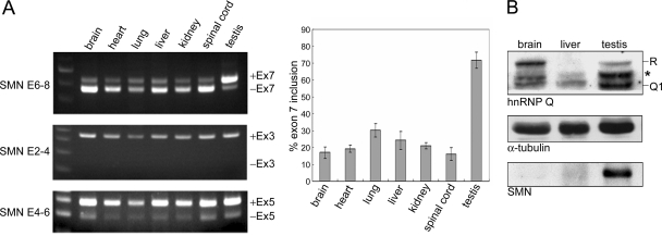
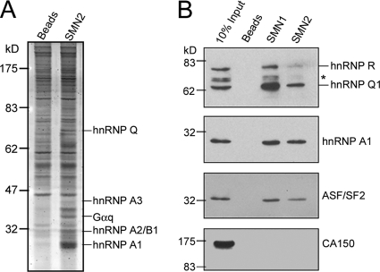
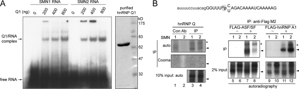
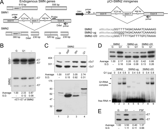
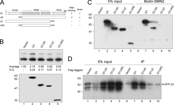
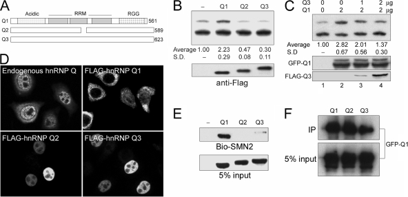
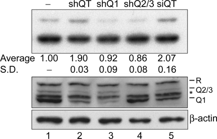
Spinal muscular atrophy (SMA) is a recessive neuromuscular disorder caused by the homozygous loss of the SMN1 gene. The human SMN2 gene has a C-to-T transition at position +6 of exon 7 and thus produces exon 7-skipping mRNAs. However, we observed an unexpectedly high level of exon 7-containing SMN2 transcripts as well as SMN protein in testis of smn(-/-) SMN2 transgenic mice. Using affinity chromatography, we identified several SMN RNA-associating proteins in mouse testis and human HeLa cells, including hnRNP Q. The major hnRNP Q isoform, Q1, directly bound SMN exon 7 in the vicinity of nucleotide +6. Overexpression of hnRNP Q1 promoted the inclusion of exon 7 in SMN2, probably by activating the use of its upstream 3' splice site. However, the minor isoforms Q2/Q3 could antagonize the activity of hnRNP Q1 and induced exon 7 exclusion. Intriguingly, enhanced exon 7 inclusion was also observed upon concomitant depletion of three hnRNP Q isoforms. Thus, differential expression of hnRNP Q isoforms may result in intricate control of SMN precursor mRNA splicing. Here, we demonstrate that hnRNP Q is a splicing modulator of SMN, further underscoring the potential of hnRNP Q as a therapeutic target for SMA.


Figures

FIG. 1.
FIG. 1.
SMN expression in different tissues of smn−/− SMN2 mice. (A) Total RNA was prepared from smn−/− SMN2 mouse tissues and analyzed by RT-PCR using specific primers (see Materials and Methods). The reaction products were then subjected to agarose gel electrophoresis. Representative data are shown at left. PCR product sizes in SMN exons 6 to 8 (E6-8), exons 2 to 4 (E2-4), and exons 4 to 6 (E4-6) were as follows: exon 7 inclusion (+Ex7), 419 bp; exon 7 exclusion (−Ex7), 367 bp; +Ex3, 350 bp; −Ex3, 149 bp; +Ex5, 288 bp; −Ex5, 192 bp. The bar graph shows exon 7 inclusion efficiency (see Materials and Methods). (B) Immunoblotting shows SMN, hnRNP Q/R, and α-tubulin protein levels in the indicated tissue cell lysates of smn−/− SMN2 mice. Anti-hnRNP Q cross-reacted with hnRNP R proteins. The asterisk may represent hnRNP Q2/3 or hnRNP R isoforms or degraded fragments.
FIG. 2.
FIG. 2.
hnRNP Q association with SMN exon 7 RNA. (A) Affinity selection of SMN2 exon 7-associated proteins was performed by incubation of the HeLa cell nuclear extract with SMN2 oligoribonucleotide-conjugated resin or empty beads. Bound samples were detected by Sypro-Ruby staining on an SDS-polyacrylamide gel. Proteins identified by MS are indicated at right. (B) HEK293 cell proteins were selected by SMN1 or SMN2 exon 7 RNA resin and detected by immunoblotting with antibodies against hnRNP Q, hnRNP A1, ASF/SF2, and CA150. Anti-hnRNP Q cross-reacted with hnRNP R. The asterisk represents hnRNP Q2/3 or potential degraded fragments or isoforms of hnRNP R.
FIG. 3.
FIG. 3.
hnRNP Q1 directly binds SMN exon 7 nearby nucleotide +6. (A) Coomassie blue staining shows the purity of recombinant His-tagged hnRNP Q1 (right panel). His-tagged hnRNP Q1 was incubated with 5′ end-labeled SMN1 or SMN2 RNA oligonucleotide. The reaction were analyzed by electrophoresis on a nondenaturing polyacrylamide gel. (B) SMN RNA oligonucleotides containing a single [32P]phosphate prior to nucleotide +6 were used for cross-linking. Lysates were prepared from HEK293 cells that were nontransfected (lanes 1 to 4) or transfected with an empty vector (lanes 5, 6, 9, and 10), or the vector expressing FLAG-tagged ASF/SF2 (lanes 7 and 8) or hnRNP A1 (lanes 11 and 12). These lysates were then incubated with SMN RNA for UV cross-linking. After RNase digestion, immunoprecipitation was performed using anti-mouse immunoglobulin (lanes 1 and 2, control antibody [Con Ab]), anti-hnRNP Q (lanes 3 and 4), or anti-FLAG (lanes 5 to 12). Input and precipitated proteins were visualized by autoradiography. Immunoprecipitated (IP) hnRNP Q is also shown by Coomassie (Cooma) blue staining. Arrowheads indicate hnRNP Q, FLAG-ASF/SF2, and FLAG-hnRNP A1. Radiolabeled signals (asterisks) might result from incomplete RNase digestion.
FIG. 4.
FIG. 4.
hnRNP Q1 promotes inclusion of exon 7 in SMN2. (A) The left diagram shows endogenous SMN1 and SMN2 transcripts and their RT-PCR products amplified using primers as indicated by arrows. Note that only SMN2 contains a DdeI restriction site. Southern blotting was performed using the probe as indicated. The right diagram shows the pCI-SMN2 reporter used in the in vivo splicing assay. Expression of the SMN transcript was driven by the cytomegalovirus (CMV) promoter. (B) HEK293 cells were transfected with GFP (G), GFP-hnRNP Q1 (Q1), or empty (−) expression vector. RT-PCR and following analysis are as described in panel A. E7, exon 7. (C) The pCI-SMN2 reporter was cotransfected into HEK293 cells along with expression vector encoding GFP (G) or GFP-ASF/SF2, SRp30c, or hnRNP Q1. RT-PCR was performed to detect SMN transcripts produced from the reporters described in panel A. Ex7, exon 7. (D) Similar to panel C, the wild-type pCI-SMN2 and its ug and r20 mutants were each cotransfected with expression vector encoding GFP or GFP-hnRNP Q1 (upper panel). His-tagged hnRNP Q1 was incubated with 32P-labeled wild-type or mutant SMN RNA oligonucleotide, and the resulting complexes were fractionated on a nondenaturing polyacrylamide gel (lower panel). The asterisk may represent a dimerized hnRNP Q1/RNA complex The SMN-ug RNA may form a secondary structure or weakly associate with hnRNP Q1 (lanes 4 to 6). (E) The pCI-SMN2 reporter was cotransfected with GFP (G), GFP-hnRNP Q1 (Q1), empty (−) or myc-PSF (PSF) expression vector into human SMA fibroblasts. Calculation of the relative change in exon 7 (E7) inclusion, shown in panels B to D, is described in Materials and Methods, and immunoblotting was performed using anti-GFP, as described for panel C. SD, standard deviation.
FIG. 5.
FIG. 5.
The C-terminal domain of hnRNP Q1 is essential for SMN2 exon 7 inclusion perhaps by its ability to bind SMN2 exon 7 and form homodimers. (A) Diagram shows hnRNP Q1 and truncated versions used in the splicing assay. The RNA binding and dimerization abilities of hnRNP Q isoforms are summarized; the asterisk indicates weak binding. (B) The pCI-SMN2 reporter was cotransfected with vector expressing FLAG-tagged full-length or truncated hnRNP Q1 proteins (Q1ΔN, Q1ΔC, or Q1ΔNC) into HEK293 cells. Splicing assay, quantitative analysis, and immunoblotting were as described in the legend of Fig. 4. SD, standard deviation. (C) HEK293 cells containing overexpressed FLAG-tagged full-length or truncated hnRNP Q1 were incubated with SMN2 oligoribonucleotide-conjugated resin. SMN RNA-selected proteins were analyzed by immunoblotting with anti-FLAG. Cells transfected with an empty vector served as a control (Vector). (D) GFP-hnRNP Q1 was overexpressed alone (Vector) or coexpressed with FLAG-tagged full-length or truncated hnRNP Q1 in HEK293 cells. Immunoprecipitation (IP) was performed in the presence of RNase using anti-FLAG, and immunoblotting used anti-GFP.
FIG. 6.
FIG. 6.
The hnRNP Q isoforms have opposing activity in alterative splicing of SMN2. (A) Diagram shows three hnRNP Q isoforms. (B) The pCI-SMN2 reporter was cotransfected with an empty vector or vector encoding FLAG-hnRNP Q1, Q2, or Q3. Splicing assay, quantitative analysis, and immunoblotting were performed as in described in the legend of Fig. 4. Ex7 represents exon 7. (C) The pCI-SMN2 reporter was cotransfected with the GFP-hnRNP Q1 expression vector alone or together with the vector encoding FLAG-hnRNP Q3 (lanes 3 and 4). Lane 1 shows a mock transfection. Immunoblotting was performed using anti-GFP and anti-FLAG to detect hnRNP Q1 and Q3, respectively. (D) Indirect immunofluorescence was performed using anti-hnRNP Q in nontransfected HeLa cells or using anti-FLAG in cells that transiently expressed FLAG-hnRNP Q1, Q2, or Q3. (E) HEK293 cell lysates containing overexpressed FLAG-hnRNP Q isoform were subjected to biotinylated-SMN2 (Bio-SMN2) RNA affinity chromatography as in described in the legend of Fig. 5C. (F) GFP-hnRNP Q1 was coexpressed with each of the FLAG-hnRNP Q isoforms in HEK293 cells. Subsequent immunoprecipitation and immunoblotting were performed as in described in the legend of Fig. 5D. SD, standard deviation.
FIG. 7.
FIG. 7.
Downregulation of hnRNP Q proteins enhances exon 7 inclusion. HEK293 cells were transfected with empty pRS vector (−) or pRS-shQT, -shQ1, or -shQ2/3 or siRNA mixture siQT. Immunoblotting (bottom) was performed to show the expression level of hnRNP Q/R isoforms and β-actin. The asterisk may represent hnRNP R isoforms or degraded fragments. The splicing assay (top) and quantitative analysis were performed as described in the legend of in Fig. 4. SD, standard deviation.

Similar articles

Cited by

References

    1. Abdul-Manan, N., S. M. O'Malley, and K. R. Williams. 1996. Origins of binding specificity of the A1 heterogeneous nuclear ribonucleoprotein. Biochemistry 353545-3554. - PubMed
    1. Black, D. L. 2003. Mechanisms of alternative pre-messenger RNA splicing. Annu. Rev. Biochem. 72291-336. - PubMed
    1. Cartegni, L., and A. R. Krainer. 2002. Disruption of an SF2/ASF-dependent exonic splicing enhancer in SMN2 causes spinal muscular atrophy in the absence of SMN1. Nat. Genet. 30377-384. - PubMed
    1. Chang, J. G., H. M. Hsieh-Li, Y. J. Jong, N. M. Wang, C. H. Tsai, and H. Li. 2001. Treatment of spinal muscular atrophy by sodium butyrate. Proc. Natl. Acad. Sci. USA 989808-9813. - PMC - PubMed
    1. Elvira, G., S. Wasiak, V. Blandford, X. K. Tong, A. Serrano, X. Fan, M. del Rayo Sanchez-Carbente, F. Servant, A. W. Bell, D. Boismenu, J. C. Lacaille, P. S. McPherson, L. DesGroseillers, and W. S. Sossin. 2006. Characterization of an RNA granule from developing brain. Mol. Cell. Proteomics 5635-651. - PubMed

Publication types

MeSH terms

LinkOut - more resources