. 2008 Dec 11;27(58):7235-47.
doi: 10.1038/onc.2008.337. Epub 2008 Sep 15.

Ligand-dependent ubiquitination of Smad3 is regulated by casein kinase 1 gamma 2, an inhibitor of TGF-beta signaling

Affiliations

Ligand-dependent ubiquitination of Smad3 is regulated by casein kinase 1 gamma 2, an inhibitor of TGF-beta signaling

X Guo et al. Oncogene. .

Abstract

Transforming growth factor-beta (TGF-beta) elicits a variety of cellular activities primarily through a signaling cascade mediated by two key transcription factors, Smad2 and Smad3. Numerous regulatory mechanisms exist to control the activity of Smad3, thereby modulating the strength and specificity of TGF-beta responses. In search for potential regulators of Smad3 through a yeast two-hybrid screen, we identified casein kinase 1 gamma 2 (CKIgamma2) as a novel Smad3-interacting protein. In mammalian cells, CKIgamma2 selectively and constitutively binds Smad3 but not Smad1, -2 or -4. Functionally, CKIgamma2 inhibits Smad3-mediated TGF-beta responses including induction of target genes and cell growth arrest, and this inhibition is dependent on CKIgamma2 kinase activity. Mechanistically, CKIgamma2 does not affect the basal levels of Smad proteins or activity of the receptors. Rather, CKIgamma2 preferentially promotes the ubiquitination and degradation of activated Smad3 through direct phosphorylation of its MH2 domain at Ser418. Importantly, mutation of Ser418 to alanine or aspartic acid causes an increase or decrease of Smad3 activity, respectively, in the presence of TGF-beta. CKIgamma2 is the first kinase known to mark activated Smad3 for destruction. Given its negative function in TGF-beta signaling and its reported overexpression in human cancers, CKIgamma2 may act as an oncoprotein during tumorigenesis.


Figures

Figure 1
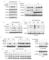
Figure 1. CKIγ2 constitutively and selectively interacts with Smad3
(a) 293T cells were transfected with the indicated constructs. Cells were lysed in universal lysis buffer supplemented with protease and phosphatase inhibitors (ULB+) at 24 h post-transfection and anti-HA immunoprecipitation was performed. 2SD, S423/425D; KD, kinase-dead; WCL, whole-cell lysate. (b) The Flag-tagged, full-length Smad constructs were co-expressed with GFP-CKIγ2(WT). Anti-Flag immunoprecipitation was performed as in (a). The arrowhead indicates the heavy chain (hc) of the Flag antibody (M2). (c) Parental HaCaT cells were pretreated with 10 µM MG-132 for 1 h before treatment with or without 100 pm TGF-β for another 2 h. Endogenous CKIγ2 was precipitated with a goat polyclonal anti-CKIγ2 antibody (C-20), and endogenous Smad proteins were blotted with a monoclonal anti-Smad1/2/3 antibody. Co-precipitated Smad3 is indicated by an arrow. An irrelevant goat polyclonal antibody (IgG) was used as a negative control. (d) Wild-type Smad3 and CKIγ2 were co-expressed in MEFs for 20h. Cells were then treated with 100pm TGF-β for the indicated time course and then lysed for anti- Flag immunoprecipitation. CKIγ2, casein kinase 1 gamma 2; HA, hemagglutinin; MEFs, mouse embryonic fibroblasts; TGF-β, transforming growth factor-beta.
Figure 2
Figure 2. CKIγ2 inhibits Smad3-mediated transcription
(a) Validation of CKIγ2 shRNAs. Flag-CKIγ2 (1µg) was co-transfected into 293T cells with the indicated pSuperRetro plasmids (pSR, 3 µg). pSR-GL2 contains the shRNA against luciferase and was used as a non-targeting control. Cells were lysed at 24 h post-transfection and total cell lysates were blotted for Flag-CKIγ2. γ-Tubulin was used as the loading control in this and most later experiments. (b) SBE-Lux luciferase assay in HepG2 cells. The luciferase reporter (0.5 µg) was co-transfected with the indicated pSuperRetro plasmids (3 µg) before TGF-β (100 pm) or vehicle was added. Data were presented as average ± s.d. (c and d). Similar luciferase assays in HepG2 cells using the MBE-Lux reporter (c) or the ARE-Luc reporter (d, together with an equal amount of FoxH1). Cells were co-transfected with the reporter construct (1 µg) and the indicated pSuperRetro plasmids (4 µg of pSR-GL2 or a combination of pSR-CKIγ2-R1 and -R2, 2 mg each). Twenty-four hours later, cells were treated with TGF-β (100 pm) or the TβRI inhibitor, SB431542 (10 µm), for another 16 h before analysis. (e) 3TP-Luc luciferase assays in HaCaT stable lines (see Materials and methods). Cells were transiently transfected with the reporter construct (1 µg) and treated with or without TGF-β (100 pm). Note that overexpressed CKIγ2 was transiently knocked down in the CKIγ2-WT cells (lane 3) by three consecutive transfections with pSuper-CKIγ2. CKIγ2 protein levels in each HaCaT line are shown (bottom). Actin was blotted to show equal loading of lysates. CKIγ2, casein kinase 1 gamma 2; shRNA, short hairpin-type RNA; TGF-β, transforming growth factor-beta; TβRI, TGF-β receptor I.
Figure 3
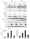
Figure 3. CKIγ2 impairs Smad3-dependent TGF-β activity
(a) Cell proliferation assays in HaCaT cells. The indicated HaCaT stable lines were treated with different concentrations of TGF-β for 24 h. The amounts of incorporated 3H-thymidine in untreated cells were normalized to 100%. (b) Downregulation of endogenous c-myc protein was measured in the HaCaT stable lines following TGF-β treatment as indicated (100 pm). Equal loading of samples was confirmed by anti-actin blotting (data not shown). (c and d) Induction of endogenous p21 protein by TGF-β in the HaCaT stable lines. Note that a lower concentration of TGF-β (10 pm) was used to manifest the enhanced responsiveness of the CKIγ2 knockdown cells (d). (e) Wound-healing assays in the HaCaT stable lines treated with SB431542 (10 µm) or TGF-β (100 pm). Asterisk (*), P≤0.01. (f) Wild-type or kinase-dead CKIγ2 was transiently expressed in MEFs. After treatment with TGF-β (50 pm), whole-cell lysates (in ULB+) were blotted for the indicated proteins. CKIγ2, casein kinase 1 gamma 2; MEFs, mouse embryonic fibroblasts; TGF-β, transforming growth factor-beta.
Figure 4
Figure 4. CKIγ2 promotes the proteasomal degradation of activated Smad3
(a) CKIγ2 does not regulate the basal levels of Smad proteins. HaCaT stable lines were treated with SB431542 (10 µm) overnight, and endogenous Smad2, -3 and -4 were examined by western blotting. (b) The HaCaT lines were transiently stimulated with TGF-β (50pm) as indicated. C-terminal phosphorylation of endogenous Smad2 and Smad3 was measured. (c) HaCaT cells were treated with TGF-β (100 pm) for the indicated time courses in the absence (left) or presence (right) of 20 µm MG-132. The level of activated Smad3 was shown. (d) HaCaT cells were treated with TGF-β (100 pm) for the indicated time courses and the total levels of endogenous Smad2 and Smad3 were determined. (e) MEFs were transfected with or without wild-type CKIγ2 before incubation with TGF-β (50pm) for the indicated time course. C-terminal phosphorylation of endogenous Smad2 and Smad3 was measured. (f) HaCaT cells were pretreated with MG-132 (7.5 µM) for 1 h before the treatment of SB431542 (10 µm, ‘−’) or TGF-β (100 pm, ‘+’) for another 3 h. Cells were harvested in SDS lysis buffer (see Materials and methods), endogenous Smad3 was immunoprecipitated and ubiquitinated species of Smad3 was analysed by anti-ubiquitin blotting. (g) MEFs expressing either vector control or wild-type CKIγ2 were treated with SB431542 (10 mm, ‘−’) or TGF-β (100 pm, ‘+’) for 2 h in the presence of 20 µm MG-132. The interaction between endogenous Smad3 and β-TrCP was determined by co-immunoprecipitation assays. CKIγ2, casein kinase 1 gamma 2; MEFs, mouse embryonic fibroblasts; TGF-β, transforming growth factor-beta.
Figure 5
Figure 5. CKIγ2 phosphorylates Smad3 at Ser418
(a) CKIγ2 in vitro kinase assay. Flag-tagged CKIγ2 (WT or KD) immunoprecipitated from 293T cell lysates as well as bacterially purified His-CKIγ2 were individually incubated with α-casein (positive control) or GST-Smad3(WT) in the presence of [32P]- γ-ATP. Phosphorylated proteins were visualized by autoradiography. Note that CKIγ2 underwent significant autophosphorylation. (b) His-CKIγ2 was incubated with GST alone, GST-S3C(WT) or GST-S3C( S418A) in a similar kinase assay as in (a). Equal loading of protein substrates was confirmed by Coomassie Blue staining (data not shown). CKIγ2 does not phosphorylate the GST moiety. (c) The indicated Flag-tagged Smad3 fragments were co-expressed with either a vector control or wild-type CKIγ2 in MEFs without inhibition of proteasomal function. Total cell lysates were analysed for the levels of Flag-S3NL (MH1 + Linker) and Flag-S3C (MH2). CKIγ2, casein kinase 1 gamma 2; GST, glutathione S-transferase; KD, kinase-dead; MEFs, mouse embryonic fibroblasts; WT, wild-type.
Figure 6
Figure 6. Ser418 phosphorylation regulates Smad3 stability and activity following TGF-β treatment
(a) MEFs expressing Smad3(WT or S418D) were treated with TGF-β (50 pm) for the indicated time course in the absence or presence of 20 µm MG-132. Induction of Smad3 target genes and Smad3 tail phosphorylation are shown. (b) MEFs were transfected with Smad3(WT or S418A) and CKIγ2 (or a vector control). After TGF-β (50 pm) treatment for the indicated time course, induction of Smad3 target genes and Smad3 tail phosphorylation were analysed. (c) Smad3-null MEFs were co-transfected with the SBE-Lux reporter and a vector control or the indicated Smad3 variants. Luciferase activity was measured after treatment with SB431542 or 100 pm TGF-β. (d) HepG2 cells expressing a vector control or the indicated Smad3 variants were treated with or without TGF-β (200 pm) for 16 h. The extent of growth inhibition (with reference to untreated cells) under each condition is shown. MEFs, mouse embryonic fibroblasts; TGF-β, transforming growth factor-beta; WT, wild-type.
Figure 7
Figure 7. The Smad3 ‘life cycle’ and regulators of Smad3 ubiquitination
Smad3 ubiquitination has important functions in determining both the intensity and the duration of TGF-β signals. Steady-state Smad3 is constantly degraded by the proteasome under the regulation of Axin, GSK3-β and probably CKIα. On the other hand, proteasomal degradation of activated Smad3 requires CKIγ2 phosphorylation, which may happen in the cytoplasm or in the nucleus or both. CKIγ2, casein kinase 1 gamma 2; TGF-β, transforming growth factor-beta.

Similar articles

Cited by

References

    1. Bierie B, Moses HL. Tumour microenvironment: TGFbeta: the molecular Jekyll and Hyde of cancer. Nat Rev Cancer. 2006;6:506–520. - PubMed
    1. Davidson G, Wu W, Shen J, Bilic J, Fenger U, Stannek P, et al. Casein kinase 1 gamma couples Wnt receptor activation to cytoplasmic signal transduction. Nature. 2005;438:867–872. - PubMed
    1. Dennler S, Huet S, Gauthier JM. A short amino-acid sequence in MH1 domain is responsible for functional differences between Smad2 and Smad3. Oncogene. 1999;18:1643–1648. - PubMed
    1. Dunn NR, Koonce CH, Anderson DC, Islam A, Bikoff EK, Robertson EJ. Mice exclusively expressing the short isoform of Smad2 develop normally and are viable and fertile. Genes Dev. 2005;19:152–163. - PMC - PubMed
    1. Fukuchi M, Imamura T, Chiba T, Ebisawa T, Kawabata M, Tanaka K, et al. Ligand-dependent degradation of Smad3 by a ubiquitin ligase complex of ROC1 and associated proteins. Mol Biol Cell. 2001;12:1431–1443. - PMC - PubMed

Publication types

MeSH terms