. 2008 Oct;15(10):1109-18.
doi: 10.1038/nsmb.1486. Epub 2008 Sep 14.

Activation of tyrosine kinases by mutation of the gatekeeper threonine

Affiliations

Activation of tyrosine kinases by mutation of the gatekeeper threonine

Mohammad Azam et al. Nat Struct Mol Biol. 2008 Oct.

Abstract

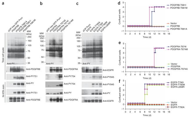
Protein kinases targeted by small-molecule inhibitors develop resistance through mutation of the 'gatekeeper' threonine residue of the active site. Here we show that the gatekeeper mutation in the cellular forms of c-ABL, c-SRC, platelet-derived growth factor receptor-alpha and -beta, and epidermal growth factor receptor activates the kinase and promotes malignant transformation of BaF3 cells. Structural analysis reveals that a network of hydrophobic interactions-the hydrophobic spine-characteristic of the active kinase conformation is stabilized by the gatekeeper substitution. Substitution of glycine for the residues constituting the spine disrupts the hydrophobic connectivity and inactivates the kinase. Furthermore, a small-molecule inhibitor that maximizes complementarity with the dismantled spine (compound 14) inhibits the gatekeeper mutation of BCR-ABL-T315I. These results demonstrate that mutation of the gatekeeper threonine is a common mechanism of activation for tyrosine kinases and provide structural insights to guide the development of next-generation inhibitors.


Figures

Figure 1
Figure 1
Sequence conservation and structural features of the gatekeeper residue threonine in tyrosine kinases and activation of kinase activity by gatekeeper residue mutation. (a) Sequence alignment of the kinase domain hinge region and conservation of the gatekeeper residue in v-SRC, c-SRC, c-ABL and several receptor tyrosine kinases. (b) Structural alignment of inactive c-SRC (cyan; PDB 1Y57) and c-ABL (gray; PDB 2G2I) showing the conformational similarities; the kinase hinge region is marked by the arrow. (c) An enlarged view of the ABL active site bound to PD166326 (above; PDB 1OPK) and an ATP analog (below; PDB 2G2F), showing the specific interaction of PD166326 but not ATP with the gatekeeper threonine. (d) Immunoblot analysis of HEK293T cells expressing BCR-ABL and different variants of c-ABL. Above, total cell lysates probed with anti-phosphotyrosine antibody (anti-PY). The blot was stripped and reprobed with anti-ABL. Below, immunoblots of Ni-NTA–purified histidine-tagged c-ABL proteins probed with anti-PY, followed by stripping and reprobing with anti-ABL antibody. (e) Immunoblot analysis of HEK293T cells expressing different c-SRC kinase variants. Above, total cell lysates probed with anti-PY. The blots were stripped and reprobed with anti-SRC, anti-phosphotyrosine-416 (anti-SRC-PY416) and anti-phosphotyrosine-527 (anti-SRC-PY527) antibodies. (f) Immunoblot of Ni-NTA–purified c-SRC kinase variants probed with anti-PY followed by stripping and reprobing as described in e. MW, molecular weight.
Figure 2
Figure 2
Kinase activation and BAF3 cellular transformation by gatekeeper mutants of SRC and ABL. (a) Immunoblot analysis of BaF3 cells expressing BCR-ABL and gatekeeper mutants of c-ABL. Above, total cell lysates probed with anti-phosphotyrosine antibody (anti-PY). Blots were stripped and reprobed with anti-ABL. Below, immunoblots of Ni-NTA–purified histidine-tagged c-ABL proteins probed with anti-PY, followed by stripping and reprobing with anti-ABL antibody. MW, molecular weight. (b) Immunoblot analysis of BaF3 cells expressing different c-SRC kinase variants. Above, total cell lysates probed with anti-PY. Blots were stripped and reprobed as described in Figure 1e. (c) Immunoblot of Ni-NTA–purified histidine-tagged c-SRC proteins probed with anti-PY. Blots were stripped and reprobed as in Figure 1e. (d) Cell-proliferation assay of BaF3 cells expressing BCR-ABL and different constructs of ABL variants. Cells were plated in quadruplicate in 96-well plates at a density of 5,000 cells per well in the absence of IL-3, and scored when the wells became confluent. (e) Cell-proliferation assay of BaF3 cells expressing c-SRC kinase variants. (f) Survival of mice injected with BaF3 cells expressing BCR-ABL and gatekeeper variants of c-SRC and c-ABL. Ten mice were injected for each construct.
Figure 3
Figure 3
Kinase activation and BAF3 cellular transformation by gatekeeper residue mutation of receptor tyrosine kinases. (a) Immunoblot analysis of BaF3 cells expressing gatekeeper mutants of PDGFRB. Above, total cell lysates probed with anti-phosphotyrosine antibody (anti-PY). Blots were stripped and reprobed with anti-PDGFRB antibody and anti–PDGFRB-phosphotyrosine-751 (anti-PY751). Below, immunoblots of Ni-NTA–purified histidine-tagged PDGFRB proteins probed with anti-PY, followed by stripping and reprobing with anti-PDGFRB antibody and anti-PY751. (b) Immunoblot analysis of BaF3 cells expressing gatekeeper mutants of PDGFRA. Above, total cell lysates probed with anti-PY. Blots were stripped and reprobed with anti-PDGFRA antibody and anti–PDGFRA-phosphotyrosine-754 (anti-PY754). Below, immunoblots of Ni-NTA–purified histidine-tagged PDGFRA proteins probed with anti-PY, followed by stripping and reprobing with anti-PDGFRA antibody and anti-PY754. (c) Immunoblot analysis of BaF3 cells expressing gatekeeper mutants of EGFR. Above, total cell lysates probed with anti-PY. Blots were stripped and reprobed with anti-EGFR antibody and anti–EGFR-phosphotyrosine-845 (anti-PY845). Below, immunoblots of Ni-NTA–purified histidine-tagged EGFR proteins probed with anti-PY, followed by stripping and reprobing with anti-EGFR antibody and anti-PY845. (df) Cell-proliferation assays of BaF3 cells expressing wild-type and gatekeeper mutants of PDGFRB (d), PDGFRA (e) and EGFR (f). Cells were plated in quadruplicate in 96-well plates at a density of 5,000 cells per well in the absence of IL-3 and scored when the wells became confluent.
Figure 4
Figure 4
The active conformation of ABL is stabilized by a hydrophobic spine linking the gatekeeper threonine to the activation loop. (a) The surface projections of the hydrophobic spine that assembled during ABL kinase activation in ABL-ADP conformation (shown in blue; PDB 2G2I). The gatekeeper residue, Thr334, is shown as an orange surface. (b) The surface projections of the dismantled hydrophobic spine in the inactive kinase conformation of ABL–imatinib (PDB 1OPJ). Imatinib binds to the inactive kinase which is stabilized by the DFG-out conformation caused by dismantling of the hydrophobic spine.
Figure 5
Figure 5
The hydrophobic spine in active and inactive SRC kinases. (a) Crystal structure of chicken c-SRC-T338I bound to ATPγS. SRC-T338I is homologous to T334I in human c-SRC. Chicken SRC residues are numbered according to the human c-SRC kinase numbering. The residues Leu328, Met317, Phe408 and His387, which constitute the hydrophobic spine, are shown in blue. The gatekeeper isoleucine residue is shown in orange. The activation loop is shown in red. (b) The inactive conformation of chicken c-SRC (PDB 2SRC), colored as in a. (c) Active site of Lck kinase (PDB 1QPC) in the active state bound with AMP-PNP shown in yellow. Gatekeeper residue Thr316 and the catalytic Lys273 are shown as green surfaces. The water molecules sandwiched between the residues Thr316 and Lys273 are shown as red circles. The interactions of lysine with ANP and the catalytic Glu288 are shown; bond distances are presented in angstroms. (d) Active site of SRC-T341I kinase domain (PDB 3DQW) bound with ATPγS shown in yellow. The surfaces of the side chains for residues Ile341 and Lys298 are shown in green. Interactions of Lys298 with Glu310 and ATPγS are mapped and the bond distances are indicated in angstroms. (e) Active site of insulin receptor kinase (IRK; PDB entry 1GAG) bound with ANP shown in yellow. The surfaces of the side chains for residues Met1076 and Lys1030 are shown in green. Interactions of catalytic Lys1030 with Glu1047 and ANP are mapped, and the bond distances are indicated in angstroms.
Figure 6
Figure 6
Disruption of hydrophobic-spine assembly by mutagenesis inactivates the ABL-T334I kinase. (a) Immunoblot analysis of HEK293T cells expressing BCR-ABL and different variants of ABL-T334I (analogous to the T315I substitution in BCR-ABL). Above, total cell lysates probed with anti-phosphotyrosine antibody (anti-PY) followed by stripping and reprobing with anti-ABL antibody. (b–e) Surface projections of the hydrophobic spine in the active ABL-ANP conformation (PDB 2G2I). Isoleucine substitution enhances spine assembly (b). L320G (c), M309G (d) and F401G (e) substitutions disrupt the spine. WT, wild type.
Figure 7
Figure 7
The ATP competitive inhibitor compound 14 disrupts the hydrophobic spine and inhibits c-ABL-T334I and BCR-ABL-T315I. (a) Chemical structures of PD166326, compound 14 and imatinib. (b) Superimposed structures of PD166326, compound 14 and imatinib in the active and inactive conformations of ABL kinase, showing the physical interaction with activation loop. (c) The ABL–compound 14 cocrystal structure (PDB 2HIW) indicates that compound 14 binds to the inactive DFG-out conformation, and the trifluoromethyl group of compound 14 binds deeply into the hydrophobic pocket partially formed by the dismantled hydrophobic spine. The additional anchorage gained through this interaction may drive conformational adjustments in the presence of the bulkier isoleucine side chain. (d–f) Immunoblot analysis of lysates of BaF3 cells transformed by c-ABL-T334I (d), BCR-ABL-native (e) and BCR-ABL-T315I (f), and exposed to various concentrations of PD166326 (left) and compound 14 (right). Blots were probed with anti–phosphotyrosine-412 antibody, anti-phosphotyrosine antibody and anti-ABL antibody as indicated on the right side of each blot. Concentrations of drugs are given above each lane.

References

    1. Cohen P. Protein kinases—the major drug targets of the twenty-first century? Nat. Rev. Drug Discov. 2002;1:309–315. - PubMed
    1. Dibb NJ, Dilworth SM, Mol CD. Switching on kinases: oncogenic activation of BRAF and the PDGFR family. Nat. Rev. Cancer. 2004;4:718–727. - PubMed
    1. Sawyers C. Targeted cancer therapy. Nature. 2004;432:294–297. - PubMed
    1. Sawyers CL. Opportunities and challenges in the development of kinase inhibitor therapy for cancer. Genes Dev. 2003;17:2998–3010. - PubMed
    1. Noble ME, Endicott JA, Johnson LN. Protein kinase inhibitors: insights into drug design from structure. Science. 2004;303:1800–1805. - PubMed

Publication types

MeSH terms