. 2008 Oct;9(10):1189-97.
doi: 10.1038/ni.1654. Epub 2008 Sep 14.

Mycobacterium tuberculosis blocks crosslinking of annexin-1 and apoptotic envelope formation on infected macrophages to maintain virulence

Affiliations

Mycobacterium tuberculosis blocks crosslinking of annexin-1 and apoptotic envelope formation on infected macrophages to maintain virulence

Huixian Gan et al. Nat Immunol. 2008 Oct.

Abstract

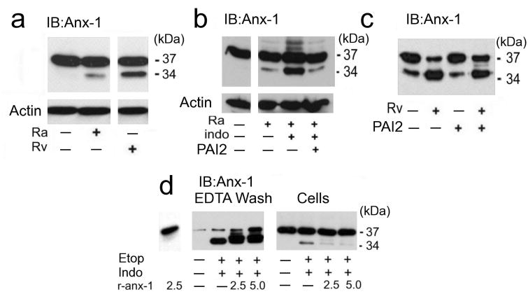
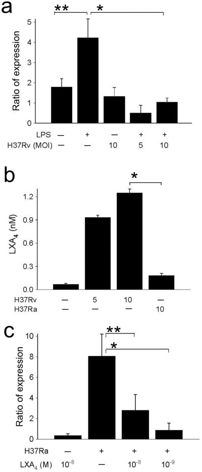
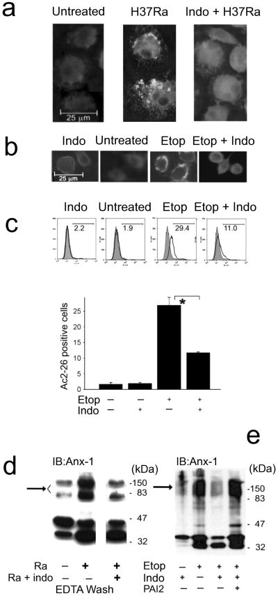
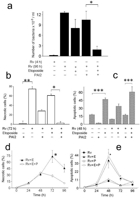
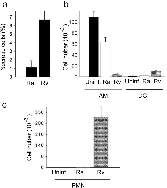
Macrophages infected with attenuated Mycobacterium tuberculosis strain H37Ra become apoptotic, which limits bacterial replication and facilitates antigen presentation. Here we demonstrate that cells infected with H37Ra became apoptotic after the formation of an apoptotic envelope on their surface was complete. This process required exposure of phosphatidylserine on the cell surface, followed by deposition of the phospholipid-binding protein annexin-1 and then transglutaminase-mediated crosslinking of annexin-1 through its amino-terminal domain. In macrophages infected with the virulent strain H37Rv, in contrast, the amino-terminal domain of annexin-1 was removed by proteolysis, thus preventing completion of the apoptotic envelope, which resulted in macrophage death by necrosis. Virulent M. tuberculosis therefore avoids the host defense system by blocking formation of the apoptotic envelope, which leads to macrophage necrosis and dissemination of infection in the lung.


Figures

Figure 1
Figure 1. Macrophages undergoing apoptosis form a surface matrix containing cross-linked annexin-1 and PAI2
(a) Immunoblot for annexin-1 or actin on lysates of human macrophages infected with Mtb strain H37Ra undergoing apoptosis and strain H37Rv undergoing necrosis (MOI 5 and 2) for 48 h. The arrow indicates high molecular weight annexin-1, which is decreased in necrotic macrophages. (b) Immunoblot for annexin-1 on lysates of H37Ra-infected (MOI 10) and uninfected macrophages surface-extracted with 2 mM EDTA in PBS. The EDTA wash and the lysed cells were immunoblotted for annexin-1. High molecular weight annexin-1 (arrow). (c,d) Immunoassays on RAW 264.7 cells treated with 5 μM etoposide (etop) or control medium for 48 h and then extracted with EDTA and immunoblotted for annexin-1 and PAI2 (c) or immunoprecipitated with annexin-1 antibody and immunoblotted for PAI2, or annexin-1 as loading control (d). (e) Immunoblot for annexin-1 or actin on lysates of RAW 264.7 cells treated with etoposide or control medium in the presence or absence of cystamine. One experiment of three is shown.
Figure 2
Figure 2. Silencing of annexin-1 abrogates apoptotic envelope formation
(a) Spectrophotometry of viable RAW 264.7 cells preincubated with vector alone or annexin-1 targeting or non-targeting siRNA and then treated with 0, 1, 5 and 10 μM etoposide for 48 h. Remaining viable cells were quantified after staining with the vital dye MTT. (*P = 0.002, n = 6).(b,c) Fluorescence microscopy for necrotic human macrophages infected with H37Ra (MOI 10, 96h) in the presence or absence of indomethacin and/or rPAI2 (b; *P = 0.07, ** P = 0.004, n = 4) or murine macrophages from wild-type and PAI2-deficient mice infected with H37Ra (MOI 10, 96 h) without or with rPAI2 (100 μg/ml) (c; *** P = 0.002, n = 3). The cells were stained with PI and counted at magnification ×100. Shown are mean percent PI+ macrophages ± s.e.(d) Immunoblot for PAI2 mRNA and 26S ribosomal RNA expression in human macrophages infected for 24 h with M. smegmatis (M. sm.), H37Ra or H37Rv (10 h). Shown is a representative from one of four experiments with similar results.
Figure 3
Figure 3. Annexin-1 cleavage is regulated by PAI2
(a-c) Immunblots of human macrophages infected with H37Rv (MOI 10, 48 h) (a,c) or H37Ra (a,b) without (a) or with (b,c) indomethacin (50 μg/ml) and/or rPAI2 (100 μg/ml), as indicated. Cell lysates were immunoblotted for annexin-1 or actin (a-c). (d) Immunoblot for annexin-1 of RAW 264.7 cells treated with etoposide (etop), indomethacin, and/or recombinant or annexin-1 (r-anx1; 2.5 or 5 μg/ml), as indicated. The cells were extracted with EDTA and the EDTA washes and cells extracts were immunoblotted. Actin staining verified equal loading (not shown). One of three experiments is shown.
Figure 4
Figure 4. Effect of virulent H37Rv on PAI2 mRNA accumulation
(a) RT-PCR for PAI2 and actin mRNAs in macrophages (5 ×10⁵ / plate) cultured with or without LPS (10 ng/ml) and infected at the MOI 10 for 24 h with virulent H37Rv. Ratio of expression refers to the n-fold-difference in PAI2 mRNA concentrations normalized to actin mRNA. (*P = 0.05, ** P = 0.01, n = 4). (b) Virulent H37Rv in contrast to avirulent H37Ra induce production of significant amounts of LXA4. Macrophages were cultured with H37Ra or H37Rv at the MOI indicated and LXA4 was measured in the supernatants after 12 h. (*P = 0.001, n = 3). (c) RT-PCR for PAI2 and actin mRNAs in macrophages infected with or without H37Ra (MOI 10, 24 h) in presence and absence of LXA4 at the concentrations indicated. Ratio of expression refers to the n-fold-difference in PAI2 mRNA concentrations normalized to actin mRNA (*P = 0.001, ** P = 0.03, n = 3).
Figure 5
Figure 5. Induction of apoptosis causes annexin-1 accumulation on the macrophage
(a,b) Fluorescence microscropy of human macrophages infected with H37Ra (MOI 10, 48 h) in the presence or absence of indomethacin (a) and RAW 264.7 cells treated with etoposide for 24 h in the presence or absence of indomethacin (b); the cells were permeabilized with digitonin and analyzed after staining with antibody to the N-terminal domain (Ac2-26) of annexin-1. One of three experiments is shown. (c) Surface expression of Ac2-26 analyzed by flow cytometry. RAW 264.7 cells treated as in b were fixed, surface-stained with Ac2-26 antibody and analyzed. Shown are histograms from a single of three experiments (top) and mean percent Ac2-26-positve cells ± s.e.(bottom, * P = 0.02, n = 3). (d,e) Immunoblots of human macrophages infected with H37Ra in the presence or absence of indomethacin and then EDTA washes of the cells were evaluated for the presence of annexin-1 (d) and RAW 264.7 cells treated as indicated with etoposide, and indomethacin and then whole cell lysates were immunoblotted for annexin-1 (e). The sample for each condition comes from 104 cells. Representative experiments of three are shown.
Figure 6
Figure 6. PAI2 protects against macrophages necrosis and contributes to expression of anti-mycobacterial activity in human macrophages infected with H37Rv
(a-e) Macrophages infected with H37Rv for the given times and with the reagents indicated. (a) Bacterial burden measured at 96 h (a, * P = 0.001, n = 6); (b) Flow cytometry for necrotic cells at 72 h as measured by PI+ cells (b, at 72 h, ** P = 0.0001, n = 5; MOI 10; * P < 0.007, n - 5); (c) TUNEL staining for apoptosis at 48 h (c,*** P = 0.015, n = 5); (d,e) ELISA photometric enzyme-immunoassay for necrosis (d) and apoptosis (e) over the same time course. (d, at 72 and 96 h, * P = 0.001, n = 3; e, P = 0.003, n = 3). One of three experiments is shown.
Figure 7
Figure 7. In vivo cytotoxicity of virulent Mtb
(a-c) Mice were challenged by tracheal instillation with 105 CFU of H37Ra or H37Rv and then cells were recovered for analysis by bronchoalveolar lavage (BAL) 9 or 14 days later. One of three experiments is shown. (a) Necrosis of BAL cells 9 days post-infection assessed by PI staining; results are expressed as the mean % PI-positive cells ± s.e.m. (P = 0.004, n = 4). (b) Resident AM and lung mDC defined as CD11blowCD11chigh and CD11bhighCD11chigh cells, respectively, quantified by flow cytometry and hemocytometer counts of total BAL cells on day 14 post-infection; the total number of either cell type in mice infected with H37Ra or H37Rv, or uninfected mice, is expressed as the mean ± s.e.m. (n = 3; infection-depleted resident AM, H37Rv versus H37Ra, P < 0.01 for differences between all three groups; the number of mDC in the lungs H37Rv versus H37Ra, P < 0.001 for Rv vs. control and H37Rv vs. H37Ra). (c) Lung neutrophils (CD11b+Gr-1+F4/80 cells) quantified by flow cytometry and hemocytometer counts of total BAL cells on day 14 post-infection expressed as the mean ± s.e.m. (n = 3; P < 0.001).

Comment in

References

    1. Leemans JC, et al. Depletion of alveolar macrophages exerts protective effects in pulmonary tuberculosis in mice. J Immunol. 2001;166:4604–4611. - PubMed
    1. Vergne I, Chua J, Singh SB, Deretic V. Cell biology of mycobacterium tuberculosis phagosome. Annu Rev Cell Dev Biol. 2004;20:367–394. - PubMed
    1. Keane J, et al. Infection by Mycobacterium tuberculosis promotes human alveolar macrophage apoptosis. Infect Immun. 1997;65:298–304. - PMC - PubMed
    1. Riendeau CJ, Kornfeld H. THP-1 cell apoptosis in response to Mycobacterial infection. Infect Immun. 2003;71:254–259. - PMC - PubMed
    1. Keane J, Remold HG, Kornfeld H. Virulent Mycobacterium tuberculosis strains evade apoptosis of infected alveolar macrophages. J Immunol. 2000;164:2016–2020. - PubMed

Publication types

MeSH terms