. 2008 Nov;88(11):1143-56.
doi: 10.1038/labinvest.2008.89. Epub 2008 Sep 15.

Integrins mediate adhesion of medulloblastoma cells to tenascin and activate pathways associated with survival and proliferation

Affiliations

Integrins mediate adhesion of medulloblastoma cells to tenascin and activate pathways associated with survival and proliferation

Paul Fiorilli et al. Lab Invest. 2008 Nov.

Abstract

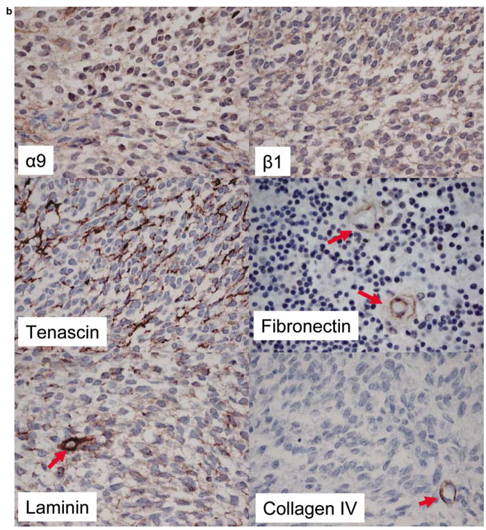
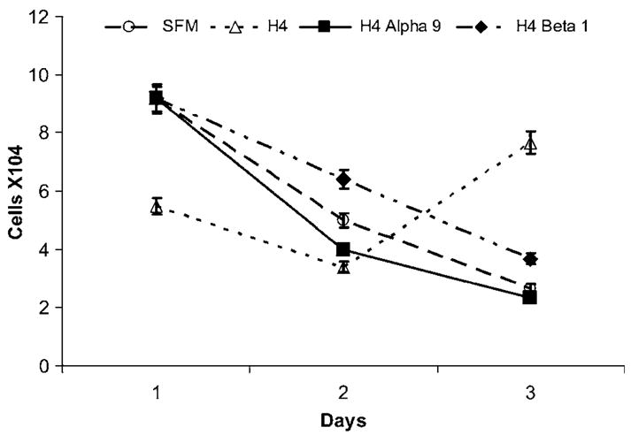
Medulloblastoma spreads by leptomeningeal dissemination rather than by infiltration that characterizes other CNS tumors, eg, gliomas. This study represents an initial attempt to identify both the molecules that mediate medulloblastoma adhesion to leptomeninges and the pathways that are key to survival and proliferation of tumor following adhesion. As a first step in molecule identification, we produced adhesion of D283 medulloblastoma cells to the extracellular matrix (ECM) of H4 glioma cells in vitro. Within this context, D283 cells preferentially expressed the alpha9 and beta1 integrin subunits; antibody and disintegrin blockade of alpha9 and beta1 binding eliminated the adhesion. The H4 ECM was enriched in tenascin, a binding partner for the alpha9beta1 integrin heterodimer. Purified tenascin-C supported D283 cell adhesion. The adhesion was blocked by antibodies to alpha9 and beta1 integrin. In vivo data were similar; immunohistochemistry of primary human medulloblastomas with leptomeningeal extension demonstrated increased expression of alpha9 and beta1 integrins as well as tenascin at the interface of brain and leptomeningeal tumor. These data suggest that tumor-cell expressions of alpha9 and beta1 integrins in combination with extracellular tenascin are necessary for medulloblastoma adhesion to the leptomeninges. As a first step in the identification of pathways that mediate survival and proliferation of tumor following adhesion, we demonstrated that adhesion to H4 ECM was associated with survival and proliferation of D283 cells as well as activation of the MAPK pathway in a growth factor deficient environment. Antibody blockade of alpha9 and beta1 integrin binding that eliminated adhesion also eliminated the in vitro survival benefit. These data suggest that adhesion of medulloblastoma to the meninges is necessary for the survival and proliferation of these tumor cells at the secondary site.


Figures

Figure 1
Figure 1
Integrin-RTK signaling. A coordinate signaling pathway from both integrins and receptor tyrosine kinases mediates cell survival and proliferation. Integrin-linked kinase (ILK) binds to the integrin β-subunit and is key to the assembly of a multiprotein complex; this multiprotein complex activates Akt. RTKs are linked via PINCH and Nck2 and initiate the Ras–MAPK pathway. (Modified from Cordes et al with permission of the authors.)
Figure 2
Figure 2
D283 medulloblastoma cells are more adherent to H4 matrix than tissue culture plastic, laminin, or fibronectin. Phase-contrast images demonstrate the minimal adhesion characteristic of D283 cells grown on tissue culture plastic (a); the degree adhesion of cells grown on laminin was similar to that of the control (b). Modest adhesion to fibronectin was observed (c). D283 cells adhered to the H4 matrix; the spreading expected to follow surface attachment in epithelial cells was also observed (d).
Figure 3
Figure 3
Adhesion of D283 cells to H4 matrix is calcium dependent. In the presence of Ca + +, 15,000 cells adhered per well. In the absence of Ca + +, a fifteenfold decrement in the binding of D283 cells to H4 matrix was observed (*P < 0.02).
Figure 4
Figure 4
D283 cells preferentially express α9 and β1 integrin subunits. α9 expression was greater than expression of the other eight α-subunits (1, 2, 3, 4, 5, 6, L, M) (*P < 0.03 for all t-tests performed). β1 expression was greater than expression of the two other β-subunits,, and the αvβ3 and αvβ5 heterodimers (*P < 4.6 × 10⁻⁵ for all t-tests performed).
Figure 5
Figure 5
Inhibition of adhesion by antibodies and disintegrins against the α9 and β1 integrin subunits. (a) Antibody to the α9-subunit inhibited 99% of adhesion and antibody to the β1-subunit inhibited 100% of adhesion to the H4 matrix (*P < 0.005 for all t-tests performed). (b) VLO5 inhibited 95% of adhesion. Echistatin inhibited only 25% of adhesion (*P < 0.04).
Figure 6
Figure 6
Tenascin is the dominate protein expressed in H4 ECM. The signal for antitenascin was more than twice as intense as that of the next largest signal (collagen IV) (*P < 0.006 for all t-tests performed).
Figure 7
Figure 7
Tenascin is the preferential substrate for α9β1 D283 adhesion. (a) More D283 cells adhere to tenascin-C than to control uncoated wells or to collagen IV (*P < 0.049). (b) Antibodies to both α9 and β1 integrin inhibit the adhesion of D283 cells to tenascin more than the adhesion to collagen IV (*P < 2 × 10⁻⁴ and 0.03, respectively).
Figure 8
Figure 8
Immunocytochemistry of α9 and β1 integrins in D283 cells. Both cell surface and cytoplasm of D283 cells labeled with (a) anti-α9 integrin and (b) VLO5.
Figure 9
Figure 9
Immunohistochemistry: increased expression of α9, β1, and tenascin in leptomeningeal implants. (a) In the normal cerebellum, staining for α9, β1, tenascin, laminin, and collagen IV was limited to sparse reactivity in vessels and meninges (black and red arrows, respectively). (All original images × 200 magnification.) (b) In primary medulloblastoma cell surface staining for α9 and β1 was present at low levels. Tenascin reactivity was observed between the cells of one tumor. There was focal laminin positivity in two tumors. Vascular staining for fibronectin, laminin, and collagen IV was observed (red arrows). (All original images × 400 magnification.) (c) In leptomeningeal implants, staining for α9, β1 (periphery, black arrows), and tenascin (interface of tumor and cerebellum red arrows) was considerable. Occasional vessels in the implants stained for laminin (black arrows). (All original images × 200 magnification.)
Figure 9
Figure 9
Immunohistochemistry: increased expression of α9, β1, and tenascin in leptomeningeal implants. (a) In the normal cerebellum, staining for α9, β1, tenascin, laminin, and collagen IV was limited to sparse reactivity in vessels and meninges (black and red arrows, respectively). (All original images × 200 magnification.) (b) In primary medulloblastoma cell surface staining for α9 and β1 was present at low levels. Tenascin reactivity was observed between the cells of one tumor. There was focal laminin positivity in two tumors. Vascular staining for fibronectin, laminin, and collagen IV was observed (red arrows). (All original images × 400 magnification.) (c) In leptomeningeal implants, staining for α9, β1 (periphery, black arrows), and tenascin (interface of tumor and cerebellum red arrows) was considerable. Occasional vessels in the implants stained for laminin (black arrows). (All original images × 200 magnification.)
Figure 10
Figure 10
D283 cells are rescued from death by adhesion to H4 matrix. D283 cells cultured in serum-free medium (SFM) in the presence of H4 matrix survived and proliferated. Absence of matrix or blockade by α9 or β1 integrin eliminated the survival benefit.
Figure 11
Figure 11
MAPK is activated following matrix adhesion of D283 medulloblastoma cells. Adhesion of D283 cells to H4 matrix resulted in threefold increase in MAPK phosphorylation (**P<0.02). At 2 hours, MAPK phosphorylation was still significantly greater than the controls (*P<0.009) (a). It did not activate Akt (b).

References

    1. MacDonald TJ, Rood BR, Santi MR, et al. Advances in the diagnosis, molecular genetics, and treatment of pediatric embryonal CNS tumors. Oncologist. 2003;8:174–186. - PubMed
    1. Zeltzer PM, Boyett JM, Finlay JL, et al. Metastasis stage, adjuvant treatment, and residual tumor are prognostic factors for medulloblastoma in children: conclusions from the Children’s Cancer Group 921 randomized phase III study. J Clin Oncol. 1999;17:832–845. - PubMed
    1. Reardon DA, Rich JN, Friedman HS, et al. Recent advances in the treatment of malignant astrocytoma. J Clin Oncol. 2006;24:1253–1265. - PubMed
    1. Mareel M, Leroy A, Bracke M. Cellular and molecular mechanisms of metastasis as applied to carcinomatous meningitis. J Neurooncol. 1998;38:97–102. - PubMed
    1. Grotzer MA, Hogarty MD, Janss AJ, et al. MYC messenger RNA expression predicts survival outcome in childhood primitive neuroectodermal tumor/medulloblastoma. Clin Cancer Res. 2001;7:2425–2433. - PubMed

Publication types

MeSH terms