

doi: 10.1038/nmeth.1258.
Epub 2008 Sep 15.
Super-resolution imaging in live Caulobacter crescentus cells using photoswitchable EYFP
Affiliations
- PMID: 18794860
- PMCID: PMC2655310
- DOI: 10.1038/nmeth.1258
Item in Clipboard
Super-resolution imaging in live Caulobacter crescentus cells using photoswitchable EYFP
Nat Methods.
2008 Nov.
Abstract
The commonly used, monomeric EYFP enabled imaging of intracellular protein structures beyond the optical resolution limit ('super-resolution' imaging) in living cells. By combining photoinduced activation of single EYFP fusions and time-lapse imaging, we obtained sub-40 nm resolution images of the filamentous superstructure of the bacterial actin protein MreB in live Caulobacter crescentus cells. These studies demonstrated that EYFP is a useful emitter for in vivo super-resolution imaging.
Figures

Reactivation of EYFP-MreB fusions in the same live C. crescentus cell. (a–l) Fluorescence images of single EYFP-MreB molecules overlaid on a reversed-contrast white-light image of the cell being examined. Initial ‘single-molecule concentration’ image showing two isolated single molecules (a). A nonemissive cell after exposure to 514-nm excitation for a few seconds (b). A short 407-nm reactivation pulse was administered after frames b, d, f, h and j in all of which no molecules are in the emissive state. Reactivated single molecules are observed in subsequent frames (c,e,g,i,k). Scale bar, 1 µm.

PALM images of EYFP-MreB in C. crescentus cells. (a,b) Banded structure in a stalked cell. (c,d) Midplane ring in a predivisional cell. Fluorescence PALM images are shown in a and c. The PALM images in b and d are the same cells as in a and c, respectively, overlaid on a reversed-contrast white-light transmission image of the cell. Scale bars, 300 nm.
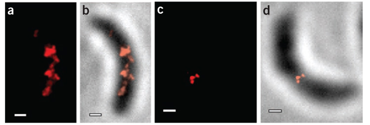
TL-PALM images of EYFP-MreB in C. crescentus cells showing fewer punctuate spots than continuous-acquisition PALM. (a,b) Quasi-helical structure in a stalked cell. (c,d) Midplane ring in a predivisional cell. Fluorescence PALM images are shown in a and c. The PALM images in b and d are the same cells as in a and c, respectively, overlaid on a reversed-contrast white-light transmission image of the cell. Scale bars, 300 nm.
References
Publication types
MeSH terms
Substances
Grants and funding
LinkOut - more resources
Full Text Sources
