. 2008 Sep;116(9):1218-25.
doi: 10.1289/ehp.11172.

Inhaled asbestos exacerbates atherosclerosis in apolipoprotein E-deficient mice via CD4+ T cells

Affiliations

Inhaled asbestos exacerbates atherosclerosis in apolipoprotein E-deficient mice via CD4+ T cells

Naomi K Fukagawa et al. Environ Health Perspect. 2008 Sep.

Abstract

Background: Associations between air pollution and morbidity/mortality from cardiovascular disease are recognized in epidemiologic and clinical studies, but the mechanisms by which inhaled fibers or particles mediate the exacerbation of atherosclerosis are unclear.

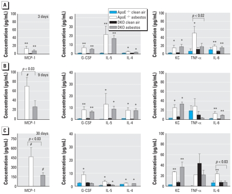
Objective and methods: To determine whether lung inflammation after inhalation of a well-characterized pathogenic particulate, chrysotile asbestos, is directly linked to exacerbation of atherosclerosis and the mechanisms involved, we exposed apolipoprotein E-deficient (ApoE(-/-)) mice and ApoE(-/-) mice crossed with CD4(-/-) mice to ambient air, NIEHS (National Institute of Environmental Health Sciences) reference sample of chrysotile asbestos, or fine titanium dioxide (TiO(2)), a nonpathogenic control particle, for 3, 9, or 30 days.

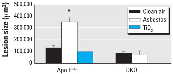
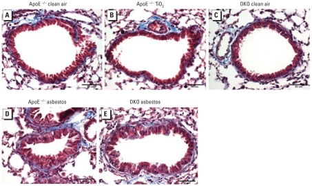
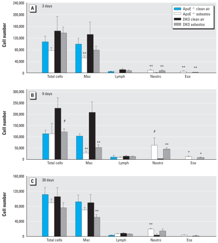
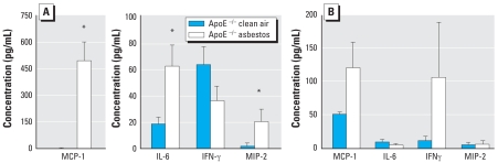
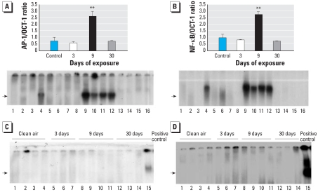
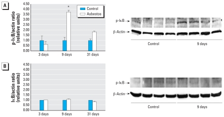
Results: ApoE(-/-) mice exposed to inhaled asbestos fibers had approximately 3-fold larger atherosclerotic lesions than did TiO(2)-exposed ApoE(-/-) mice or asbestos-exposed ApoE(-/-)/CD4(-/-) double-knockout (DKO) mice. Lung inflammation and the magnitude of lung fibrosis assessed histologically were similar in asbestos-exposed ApoE(-/-) and DKO mice. Monocyte chemoattractant protein-1 (MCP-1) levels were increased in bronchoalveolar lavage fluid and plasma, and plasma concentrations correlated with lesion size (p < 0.04) in asbestos-exposed ApoE(-/-) mice. At 9 days, activator protein-1 (AP-1) and nuclear factor-kappaB (NF-kappaB), transcription factors linked to inflammation and found in the promoter region of the MCP-1 gene, were increased in aortas of asbestos-exposed ApoE(-/-) but not DKO mice.

Conclusion: Our findings show that the degree of lung inflammation and fibrosis does not correlate directly with cardiovascular effects of inhaled asbestos fibers and support a critical role of CD4(+) T cells in linking fiber-induced pulmonary signaling to consequent activation of AP-1- and NF-kappaB-regulated genes in atherogenesis.

Keywords: AP-1; CD4+ T-cells; MCP-1; NF-κB; atherosclerosis; chrysotile asbestos; fibrosis; inflammation; knockout mice; lung.


Figures

Figure 1
Figure 1
Size (mean ± SE) of atherosclerotic lesions after 30-day exposures of ApoE−/− mice to clean air (n = 11), asbestos (n = 11), or TiO2 (n = 6), and of DKO mice to clean air (n = 8) or asbestos (n = 8). Error bars represent the variance in the average lesion area per animal. *p < 0.001.
Figure 2
Figure 2
Representative lung sections stained with Masson’s trichrome after 30-day exposures, showing inflammatory cell infiltration and fibrosis in asbestos-exposed animals. Blue indicates collagen associated with fibrosis. (A) ApoE−/− mice exposed to clean air. (B) ApoE−/− mice exposed to TiO2. (C) DKO mice exposed to clean air. (D) ApoE−/− mice exposed to asbestos. (E) DKO mice exposed to asbestos.
Figure 3
Figure 3
Differential cell counts (mean ± SE) in BALF from ApoE−/− and DKO mice after exposure for 3 days (A), 9 days (B), or 30 days (C) to clean air or asbestos. Error bars represent the variance of the averages of the respective cell counts per animal. Eos, eosinophils; Lymph, lymphocytes; Mac, macrophages; Neutro, neutrophils. *p < 0.05, **p < 0.01, and #p < 0.001, compared with respective clean-air–exposed animals (n = 5–10 animals per group per treatment).
Figure 4
Figure 4
Cytokine concentrations (mean ± SE) in BALF (A) and plasma (B) obtained from ApoE−/− mice exposed to clean air or asbestos for 30 days (n =11 per group). Error bars represent the variance of the averages of values obtained in each animal. *p < 0.04 compared with respective clean-air–exposed animals.
Figure 5
Figure 5
Cytokine and chemokine concentrations (mean ± SE) in BALF from ApoE−/− and DKO mice exposed to clean air or chrysotile asbestos for 3 days (A), 9 days (B), or 30 days (C), as analyzed by the Bio-Plex assay. Error bars represent the variance of the averages of respective values from each animal. *p < 0.05, **p < 0.01, and #p < 0.001, compared with respective clean-air–exposed animals.
Figure 6
Figure 6
Results of EMSA for NF-κB and AP-1 in aortic extracts. (A,B) DNA binding of AP-1 (A) and NF-κB (B) in aortic extracts from ApoE−/− mice exposed to clean air (control; lanes 1–4) or to chrysotile asbestos for 3 days (lanes 5–8), 9 days (lanes 9–12 ), or 30 days (lanes 13–16). Data are expressed relative to OCT-1 compound as a loading control. Values in top portions of A and B are mean ± SE; error bars represent the variance of the averages of values obtained for each animal. (C,D) DNA binding of AP-1 (C) and NF-κB (D) in aortic extracts from DKO mice exposed to clean air (lanes 1–4) or to asbestos for 3 days (lanes 5–7), 9 days (lanes 8–11), or 30 days (lanes 12–14). Lane 15 represents a positive control. Arrows denote bands for respective transcription factor binding. **p < 0.01 compared with control (n = 4 per group).
Figure 7
Figure 7
Results of Western blot analyses for phosphorylated IκB (p-IκB). (A) Western blot analyses (mean ± SE; n = 4 per group) for p-IκB/β-actin ratios in cytoplasmic extracts from aortas of ApoE−/− mice exposed to clean air (control) or chrysotile asbestos for 3, 9, or 30 days (left), and an example of a Western blot comparing clean air with 9-day exposures (right). (B) Western blot analyses (mean ± SE; n = 4 per group) for p-IκB/β-actin ratios in cytoplasmic extracts from aortas of DKO mice exposed to clean air (control) or chrysotile asbestos for 3, 9, or 30 days (left), and an example of the Western blot comparing clean air with 9-day exposures (right). Blots are not shown for 3- and 30-day exposures because there were no significant changes. *p < 0.02 compared with clean air exposure.

References

    1. BéruBé KA, Quinlan TR, Moulton G, Hemenway D, O’Shaughnessy P, Vacek P, et al. Comparative proliferative and histopathologic changes in rat lungs after inhalation of chrysotile or crocidolite asbestos. Toxicol Appl Pharmacol. 1996;137:67–74. - PubMed
    1. Brook RD, Franklin B, Cascio W, Hong Y, Howard G, Lipsett M, et al. Air pollution and cardiovascular disease: a statement for healthcare professionals from the Expert Panel on Population and Prevention Science of the American Heart Association. Circulation. 2004;109:2655–2671. - PubMed
    1. CDC (Centers for Disease Control and Prevention) Potential exposures to airborne and settled surface dust in residential areas of lower Manhattan following the collapse of the World Trade Center—New York City, November 4-December 11, 2001. Morbid Mortal Wkly Rep. 2003;52:131–136. - PubMed
    1. Craighead JE. Asbestos-associated dieases: report of the Pneumoconiosis Committee of the College of American Pathologists and the National Institute for Occupational Safety and Health. Arch Pathol Lab Med. 1982;106:543–597. - PubMed
    1. Davenport P, Tipping PG. The role of interleukin-4 and interleukin-12 in the progression of atherosclerosis in apolipoprotein E-deficient mice. Am J Pathol. 2003;163:1117–1125. - PMC - PubMed

Publication types