

Spinal angiolipoma: case report and literature review
- PMID: 18795485
- PMCID: PMC2565558
- DOI: 10.1080/10790268.2008.11760731
Spinal angiolipoma: case report and literature review
Abstract
Background/objective: Spinal angiolipoma (SAL) is an uncommon clinico-pathological entity.
Design: Single case report.
Methods: Retrospective data analysis.
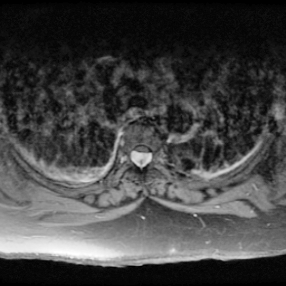
Findings: An obese woman with a 1-year history of progressive spastic paraparesis and acute deterioration underwent magnetic resonance imaging of the thoracic spine, the results of which suggested a tumor compressing the thoracic spinal cord. The histopathological examination of the completely resected tumor revealed an epidural angiolipoma.
Conclusions: This case report offers a reminder that SAL should be considered in the differential diagnosis of long-standing, slowly progressive paraparesis. It remains unclear whether an increased body mass index might be a contributing factor to the development of SAL.
Figures


References
-
- Andaluz N, Balko G, Bui H, Zuccarello M. Angiolipomas of the central nervous system. J Neuro Oncol. 2000;49((3)):219–230. - PubMed
-
- Lin JJ, Lin F. Two entities in angiolipoma. A study of 459 cases of lipoma with review of literature on infiltrating angiolipoma. Cancer. 1974;34((3)):720–727. - PubMed
-
- Fourney DR, Tong KA, Macaulay RJ, Griebel RW. Spinal angiolipoma. Can J Neurol Sci. 2001;28((1)):82–88. - PubMed
-
- Labram EK, el-Shunnar K, Hilton DA, Robertson NJ. Revisited: spinal angiolipoma—three additional cases. Br J Neurosurg. 1999;13((1)):25–29. - PubMed
-
- Pagni CA, Canavero S. Spinal epidural angiolipoma: rare or unreported. Neurosurgery. 1992;31((4)):758–764. discussion 764. - PubMed
Publication types
MeSH terms
LinkOut - more resources
Full Text Sources