. 2008 Oct 7;47(40):10649-56.
doi: 10.1021/bi801128d. Epub 2008 Sep 17.

Probing the oligomeric assemblies of pea porphobilinogen synthase by analytical ultracentrifugation

Affiliations

Probing the oligomeric assemblies of pea porphobilinogen synthase by analytical ultracentrifugation

Bashkim Kokona et al. Biochemistry. .

Abstract

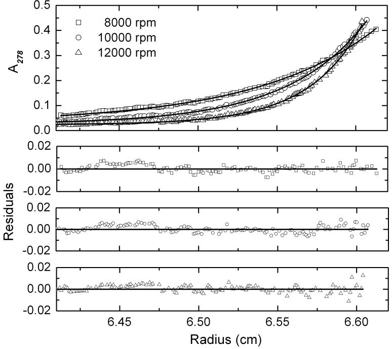
The enzyme porphobilinogen synthase (PBGS) can exist in different nonadditive homooligomeric assemblies, and under appropriate conditions, the distribution of these assemblies can respond to ligands such as metals or substrate. PBGS from most organisms was believed to be octameric until work on a rare allele of human PBGS revealed an alternate hexameric assembly, which is also available to the wild-type enzyme at elevated pH [Breinig, S., et al. (2003) Nat. Struct. Biol. 10, 757-763]. Herein, we establish that the distribution of pea PBGS quaternary structures also contains octamers and hexamers, using both sedimentation velocity and sedimentation equilibrium experiments. We report results in which the octamer dominates under purification conditions and discuss conditions that influence the octamer:hexamer ratio. As predicted by PBGS crystal structures from related organisms, in the absence of magnesium, the octameric assembly is significantly destabilized, and the oligomeric distribution is dominated largely by the hexameric assembly. Although the PBGS hexamer-to-octamer oligomeric rearrangement is well documented under some conditions, both assemblies are very stable (under AU conditions) in the time frame of our ultracentrifuge experiments.


Figures

Figure 1
Figure 1. The different oligomeric assemblies of PBGS
The PBGS equilibrium ensemble of quaternary structure forms is illustrated using homology models of pea PBGS. For most species, the asymmetric unit of the crystal structure is an asymmetric homo-dimer (3, 5, 6, 27). The hexamer and its asymmetric unit, the detached dimer (based on PDB code 1PV8) are shown in two shades of blue. The octamer and its asymmetric unit, the hugging dimer (based on PDB code 1GZG) are shown in two shades of pink. For the octamer, the dimers assemble at a 90° rotation around a central axis; for the hexamer, the dimers assemble at a 120° rotation around a central axis. The octamer contains a phylogenetically variable binding site for an allosteric magnesium ion. This ion binds to the arm-to-barrel interface that is unique to the octamer; the allosteric magnesium binding site is not present in the hexamer (3, 22).
Figure 2
Figure 2
Electrophoretic analysis of pea PBGS. A) PAGE analysis of purified pea PBGS. SDS lane 1 - molecular weight markers (weights shown in kDa); SDS lane 2 - pea PBGS (3 mg/mL) as purified (100 mM Tris-HCl, pH 8.5, 10 mM MgCl2); Native - pea PBGS (1 mg/mL) as purified. B) Native PAGE analysis of pea PBGS (1 mg/mL). lane 1 - pea PBGS as purified; lane 2 - pea PBGS dialyzed vs. 10 mM BTP pH 8.5; lane 3 - the sample shown in lane 2 with 10 mM MgCl2 added in the protein dilution step; lane 4 - the sample shown in lane 2 with 1 mM ALA added in the protein dilution step; lane 5 - the sample shown in lane 2 with 10 mM MgCl2 and 1 mM ALA added in the protein dilution step.
Figure 3
Figure 3
Sedimentation velocity analysis of PBGS in the presence and absence of Mg2+. The g(s*) distribution of s*20,w including a fit using either a single species model or a three species model is shown. Solution conditions: 34 μM C326A pea PBGS in 0.1M BTP, pH 8.5, 0.1 mM DTT, and ± 10 mM MgCl2, at 20°C; data collected at 30,000 rpm. (A) The data are shown fit with the three species model (open triangles and open circles). (B) The residuals from fitting using single-species models display a non-random distribution, suggesting an inferior fit to this model. (C) A marked improvement is observed in the residuals from fitting using-three species models.
Figure 4
Figure 4
Sedimentation equilibrium analysis of PBGS in the presence of MgCl2. SE data are shown fit with a hexamer-octamer model where the hexamer and octamer are treated as non-equilibrating mixtures. The fit shown is that from simultaneous analysis of three protein concentrations (3, 10, and 30 μM subunits) each collected at three rotor speeds. Data were collected at 8,000, 10,000, and 12,000 rpm. The fits to all of the data are provided in Figure S1 in the Supporting Information. Sample conditions: 10 μM C326A pea PBGS in 0.1 M BTP-HCl, pH 8.5, 0.1 mM DTT, and 10 mM MgCl2, at 20°C.
Figure 5
Figure 5
Sedimentation equilibrium analysis of PBGS in the absence of MgCl2. The data are shown fit with a hexamer-octamer model where the hexamer and octamer are irreversible mixtures. The fit shown is that from simultaneous analysis of three protein concentrations (3, 10, and 30 μM) each collected at three rotor speeds. Data were collected at 8,000, 10,000, and 12,000 rpm. The fits to all of the data are provided in Figure S2 in the Supporting Information. Sample conditions: 10 μM C326A pea PBGS in 0.1 M BTP-HCl, pH 8.5, and 0.1 mM DTT, at 20°C.

References

    1. Battersby AR. Tetrapyrroles: the pigments of life. Nat Prod Rep. 2000;17:507–26. - PubMed
    1. Kervinen J, Dunbrack RL, Jr., Litwin S, Martins J, Scarrow RC, Volin M, Yeung AT, Yoon E, Jaffe EK. Porphobilinogen synthase from pea: expression from an artificial gene, kinetic characterization, and novel implications for subunit interactions. Biochemistry. 2000;39:9018–29. - PubMed
    1. Breinig S, Kervinen J, Stith L, Wasson AS, Fairman R, Wlodawer A, Zdanov A, Jaffe EK. Control of tetrapyrrole biosynthesis by alternate quaternary forms of porphobilinogen synthase. Nat Struct Biol. 2003;10:757–63. - PubMed
    1. Jaffe EK, Ali S, Mitchell LW, Taylor KM, Volin M, Markham GD. Characterization of the role of the stimulatory magnesium of Escherichia coli porphobilinogen synthase. Biochemistry. 1995;34:244–51. - PubMed
    1. Frankenberg N, Erskine PT, Cooper JB, Shoolingin-Jordan PM, Jahn D, Heinz DW. High resolution crystal structure of a Mg2+-dependent porphobilinogen synthase. J Mol Biol. 1999;289:591–602. - PubMed

Publication types

MeSH terms

Substances