. 2008 Oct 7;47(40):10620-9.
doi: 10.1021/bi801157p. Epub 2008 Sep 17.

The peripheral neuropathy-linked Trembler and Trembler-J mutant forms of peripheral myelin protein 22 are folding-destabilized

Affiliations

The peripheral neuropathy-linked Trembler and Trembler-J mutant forms of peripheral myelin protein 22 are folding-destabilized

Jeffrey K Myers et al. Biochemistry. .

Abstract

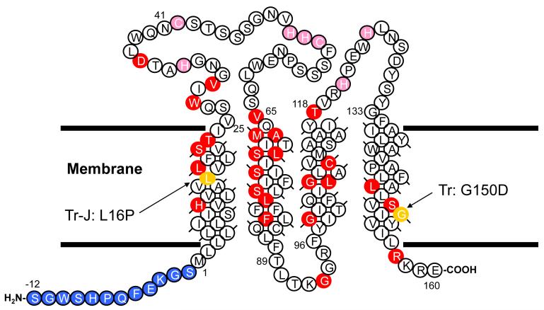
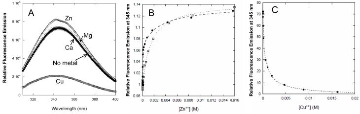
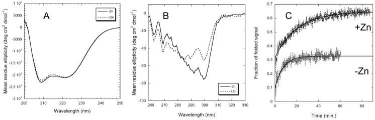
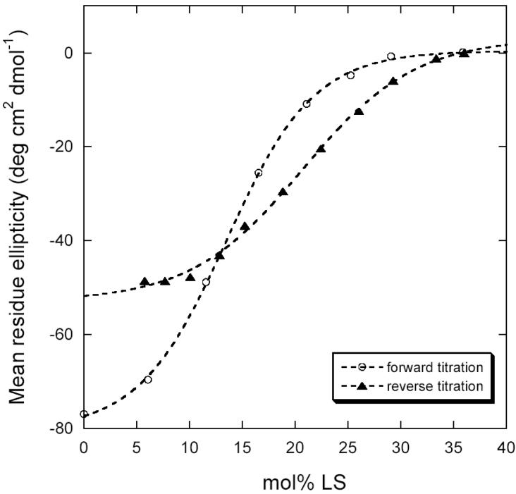
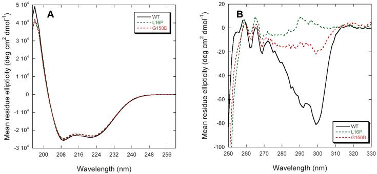
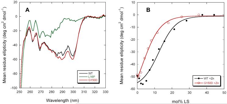
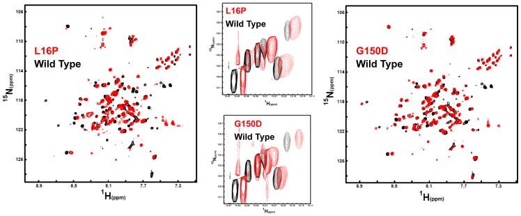
Dominant mutations in the tetraspan membrane protein peripheral myelin protein 22 (PMP22) are known to result in peripheral neuropathies such as Charcot-Marie-Tooth type 1A (CMT1A) disease via mechanisms that appear to be closely linked to misfolding of PMP22 in the membrane of the endoplasmic reticulum (ER). To characterize the molecular defects in PMP22, we examined the structure and stability of two human disease mutant forms of PMP22 that are also the basis for mouse models of peripheral neuropathies: G150D ( Trembler phenotype) and L16P ( Trembler-J phenotype). Circular dichroism and NMR spectroscopic studies indicated that, when folded, the three-dimensional structures of these disease-linked mutants are similar to that of the folded wild-type protein. However, the folded forms of the mutants were observed to be destabilized relative to the wild-type protein, with the L16P mutant being particularly unstable. The rate of refolding from an unfolded state was observed to be very slow for the wild-type protein, and no refolding was observed for either mutant. These results lead to the hypothesis that ER quality control recognizes the G150D and L16P mutant forms of PMP22 as defective through mechanisms closely related to their conformational instability and/or slow folding. It was also seen that wild-type PMP22 binds Zn(II) and Cu(II) with micromolar affinity, a property that may be important to the stability and function of this protein. Zn(II) was able to rescue the stability defect of the Tr mutant.


Figures

Figure 1
Figure 1
Amino acid sequence of human PMP22. Sites of disease-linked point mutations are marked in red, residues added to the N-terminus as part of a purification tag are shown in blue, and the locations of possible metal ion-coordinating side chains in the extracellular loops are indicated in pink. The locations of the two disease-linked mutations studied in this work are shown in gold.
Figure 2
Figure 2
PMP22 binds Cu(II) and Zn(II) at 20°C. (A) Fluorescence emission spectra of PMP22 in DPC micelles in the presence of various metal ions. The protein and metal ion concentrations were 1 μM and 1 mM, respectively. The buffer was 25 mM acetate, pH 5.5, 150 mM NaCl, 0.5 % DPC. (B) Titration with zinc acetate at pH 5.5 (open squares) and in HEPES buffer at pH 7.5 (closed circles). Single and double binding site models were fit to the data to determine dissociation constants (see Methods). Fitted Kd for Zn(II) binding were 1.4 mM at pH 5.5, and 20 μM and 3 mM at pH 7.5. (C) Titration with copper sulfate at pH 5.5. Fitted Kd were 37 μM and 1.1 mM.
Figure 3
Figure 3
The effect of Zn(II) on the structure and folding of wild type PMP22 as probed by circular dichroism. Buffer conditions were 25 mM acetate, pH 5.5, 150 mM NaCl, 0.5 % DPC, while the Zn(II) concentration was 10 mM in each experiment denoted by dotted lines. (A) Far-UV CD at 25°C, where the protein concentration in DPC micelles was 10 μM in a 0.1 cm pathlength cuvette. (B) Near UV CD at 25°C, where the protein concentration in DPC micelles was 26 μM in a 1 cm pathlength cuvette. (C) Kinetics of refolding of PMP22 at 37°C from a DPC/LS detergent mixture in the absence (black) and presence (grey) of 10 mM Zn(II). Tertiary structure formation was monitored by CD at 299 nm. Black lines are for single exponential fits of the data.
Figure 4
Figure 4
Forward (open circles) and reverse (closed triangles) titrations of wild type PMP22 in DPC micelles at 37°C with the denaturing detergent laurylsarcosine, as monitored by CD at 299 nm. Buffer was 25 mM acetate, pH 5.5, 150 mM NaCl, 0.5 % DPC with 10 mM Zn(II). Dotted lines represent fits of a two-state unfolding model (38).
Figure 5
Figure 5
Circular dichroism spectra of the Tr/G150D and TrJ/L16P mutant forms of PMP22 in DPC micelles at 25°C. (A) Far-UV CD spectra. (B) Near-UV CD spectra. Buffer conditions were 25 mM acetate, pH 5.5, 150 mM NaCl, 0.5 % DPC.
Figure 6
Figure 6
Properties of the Tr/G150D and TrJ/L16P mutant forms of PMP22 in the presence of Zn(II). (A) Near-UV CD spectra of the WT and mutants in the presence of 10 mM Zn(II). (B) Laurylsarcosine titrations followed by CD at 299nm of WT and Tr PMP22 at 37°C in the presence of Zn(II) showing the destabilization caused by the G150D mutation. Buffer was 25 mM acetate, pH 5.5, 150 mM NaCl, 0.5 % DPC.
Figure 7
Figure 7
Comparison of the NMR spectrum for WT PMP22 in TDPC micelles to the spectra of the Tr (G150D) and TrJ (L16P) mutants under identical conditions (25 mM acetate, pH 5.5, 150 mM NaCl, 0.2 % TDPC). All data were acquired at 800 MHz and 45°C. Left: The 1H,15N-TROSY spectrum of the L16P PMP22 mutant in red is overlaid on the spectrum of WT PMP22 protein in black. The L16P spectrum was collected using 128 increments and 248 scans per increment. The wild type spectrum was collected with 128 increments and 180 scans. Right: The 1H,15N-TROSY spectrum of the G150D PMP22 mutant in red is overlaid on the spectrum of WT PMP22 protein in black. Acquisition parameters for G150D were the same as those for L16P. Center: Mutant/WT TROSY spectral overlays for the indole ring 1H-15N from the protein’s 6 tryptophan residues (5 in PMP22 and 1 in the N-terminal tag).

Similar articles

Cited by

References

    1. Bronstein JM. Function of tetraspan proteins in the myelin sheath. Curr. Opin. Neurobiol. 2000;10:552–557. - PubMed
    1. Quarles RH. Myelin sheaths: glycoproteins involved in their formation, maintenance and degeneration. Cellular and Molecular Life Sciences. 2002;59:1851–1871. - PMC - PubMed
    1. Adlkofer K, Martini R, Aguzzi A, Zielasek J, Toyka KV, Suter U. Hypermyelination and demyelinating peripheral neuropathy in Pmp22-deficient mice. Nat. Genet. 1995;11:274–280. - PubMed
    1. Carenini S, Neuberg D, Schachner M, Suter U, Martini R. Localization and functional roles of PMP22 in peripheral nerves of P0-deficient mice. Glia. 1999;28:256–264. - PubMed
    1. Taylor V, Zgraggen C, Naef R, Suter U. Membrane topology of peripheral myelin protein 22. J. Neurosci. Res. 2000;62:15–27. - PubMed

Publication types