. 2009 Apr;15(4):877-85.
doi: 10.1089/ten.tea.2008.0018.

Angiogenic response to bioactive glass promotes bone healing in an irradiated calvarial defect

Affiliations

Angiogenic response to bioactive glass promotes bone healing in an irradiated calvarial defect

Ann Leu et al. Tissue Eng Part A. 2009 Apr.

Abstract

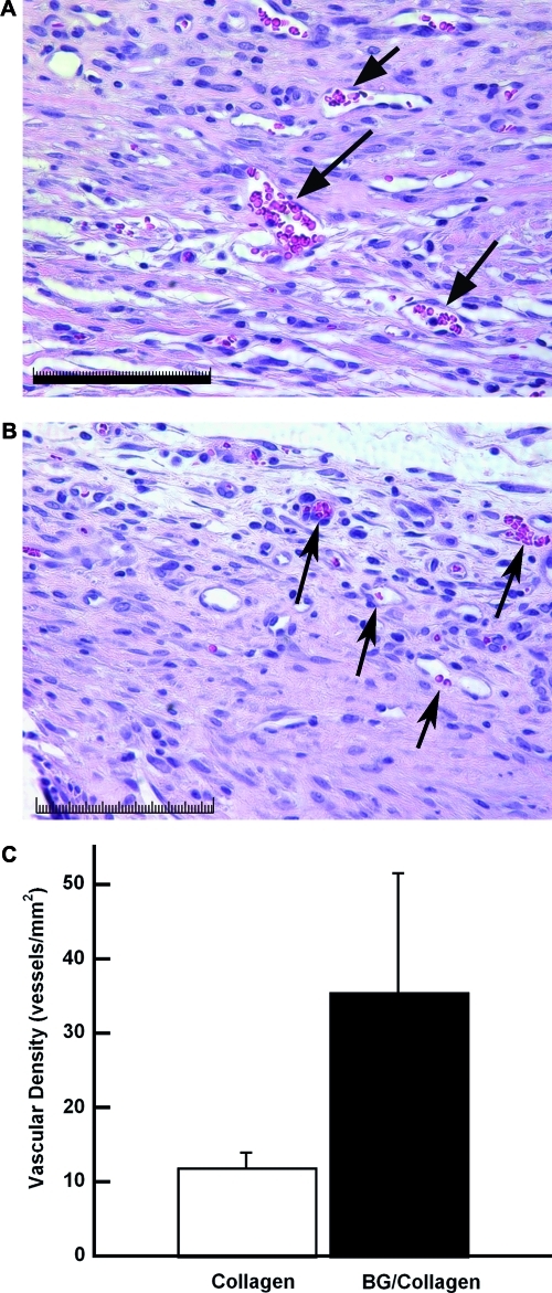
Localized radiation is an effective treatment modality for carcinomas, yet the associated reduction of the host vasculature significantly inhibits the tissue's regenerative capacity. Low concentrations of bioactive glass (BG) possess angiogenic potential, and we hypothesized that localized BG presentation would increase neovascularization and promote healing in an irradiated bone defect. An isolated calvarial region of Sprague-Dawley rats was irradiated 2 weeks before surgery. Bilateral critical-sized defects were created and immediately filled with a BG-loaded collagen sponge or an empty sponge as an internal control. Histological analysis of calvaria collected after 2 weeks demonstrated greater neovascularization within the defect in the presence of BG than with collagen alone. Noninvasive ultrasound imaging at 4 weeks detected less contrast agent in the brain below BG-treated defects than in the nearby untreated defects and images of treated defects acquired at 2 weeks. The reduced ability to detect contrast agent in BG-treated defects suggested greater attenuation of ultrasound signal due to early bone formation. Micro-computed tomography imaging at 12 weeks demonstrated significantly greater bone volume fraction within BG-treated defects than in controls. These results suggest that neovascularization induced by localized BG delivery promotes bone regeneration in this highly compromised model of bone healing and may offer an alternative approach to costly growth factors and their potential side-effects.


Figures

FIG. 1.
FIG. 1.
(A) Bilateral defects (3.5 mm in diameter) created in the calvarial region of a rat. (B) Bilateral defects containing a collagen sponge (left) and a bioactive glass–collagen sponge (right).
FIG. 2.
FIG. 2.
Two-week decalcified tissues stained with hematoxylin and eosin and treated with the bioactive glass (BG)-loaded collagen sponge (A) or collagen control (B) and imaged at 40×. Arrows denote vessels; scale bar represents 100 μm. (C) Quantification of blood vessel density of the collagen control sponges (open bar), and the BG-loaded collagen sponges (filled bar). Data are means ± standard errors of the mean. Color images available online at www.liebertonline.com/ten.
FIG. 3.
FIG. 3.
(A) Representative transverse ultrasound image of bilateral calvarial defect under contrast pulse sequencing (CPS) mode at 4 weeks before delivery of contrast agent. Defect areas are denoted by dashed lines. (B) Representative transverse ultrasound images of bilateral calvarial defect under CPS mode 5 s after the delivery of contrast agent at 2 and 4 weeks postsurgery. The defect on the right contains a bioactive glass (BG)-loaded collagen sponge; the left defect is filled with an empty collagen sponge. Defect areas are denoted by dashed lines, and perfusion regions of interest are denoted by red and yellow circles for control and BG treatment, respectively. Color scale bar represents differences in echogenicity related to perfusion and surrounding tissues. (C) Contrast intensity ratio as a function of time, defined as the ratio of contrast agent detected in the brain below control defects to BG-treated defects. Data are means ± standard errors of the mean. Color images available online at www.liebertonline.com/ten.
FIG. 4.
FIG. 4.
(A) Representative in vivo micro computed tomography (CT) image of calvaria at 12 weeks postsurgery. (B) Representative ex vivo microCT image of explanted calvaria 12 weeks postsurgery. Left defect filled with collagen sponge; right defect filled with bioactive glass (BG)-loaded collagen sponge. (C) Quantitative analysis of bone volume fraction in collagen control implants (open bar) and BG-loaded collagen implants (filled bar). Data are means ± standard errors of the mean. *P < 0.01 versus collagen control implants.
FIG. 5.
FIG. 5.
(A) Representative decalcified tissue at 12 weeks postsurgery stained with hematoxylin and eosin. Arrows denote margins of remaining defects treated using an empty collagen sponge (left) or a bioactive glass (BG)-loaded collagen sponge (right); arrowheads denote islands of mineralized nodules within BG-treated defects. Empty collagen sponges lack localized positive staining for alkaline phosphatase (B), whereas alkaline phosphatase staining of BG-treated defects confirms the presence of cells from the osteogenic lineage, as represented by reddish-brown staining (arrows) (C). 40× magnification; scale bar represents 100 μm. Color images available online at www.liebertonline.com/ten.

References

    1. American Cancer Society. Cancer Facts & Figures 2008. Atlanta: American Cancer Society; 2008.
    1. Stone H.B. Coleman C.N. Anscher M.S. McBride W.H. Effects of radiation on normal tissue: consequences and mechanisms. Lancet Oncol. 2003;4:529. - PubMed
    1. Udagawa T. Birsner A.E. Wood M. D'Amato R.J. Chronic suppression of angiogenesis following radiation exposure is independent of hematopoietic reconstitution. Cancer Res. 2007;67:2040. - PubMed
    1. Abdollahi A. Lipson K.E. Han X. Krempien R. Trinh T. Weber K.J. Hahnfeldt P. Hlatky L. Debus J. Howlett A.R. Huber P.E. SU5416 and SU6668 attenuate the angiogenic effects of radiation-induced tumor cell growth factor production and amplify the direct anti-endothelial action of radiation in vitro. Cancer Res. 2003;63:3755. - PubMed
    1. Okunieff P. Wang X. Rubin P. Finkelstein J.N. Constine L.S. Ding I. Radiation-induced changes in bone perfusion and angiogenesis. Int J Radiat Oncol Biol Phys. 1998;42:885. - PubMed

Publication types

LinkOut - more resources