. 2008 Nov;107(4):1091-101.
doi: 10.1111/j.1471-4159.2008.05687.x. Epub 2008 Sep 15.

Tissue-type plasminogen activator requires a co-receptor to enhance NMDA receptor function

Affiliations

Tissue-type plasminogen activator requires a co-receptor to enhance NMDA receptor function

Andre L Samson et al. J Neurochem. 2008 Nov.

Abstract

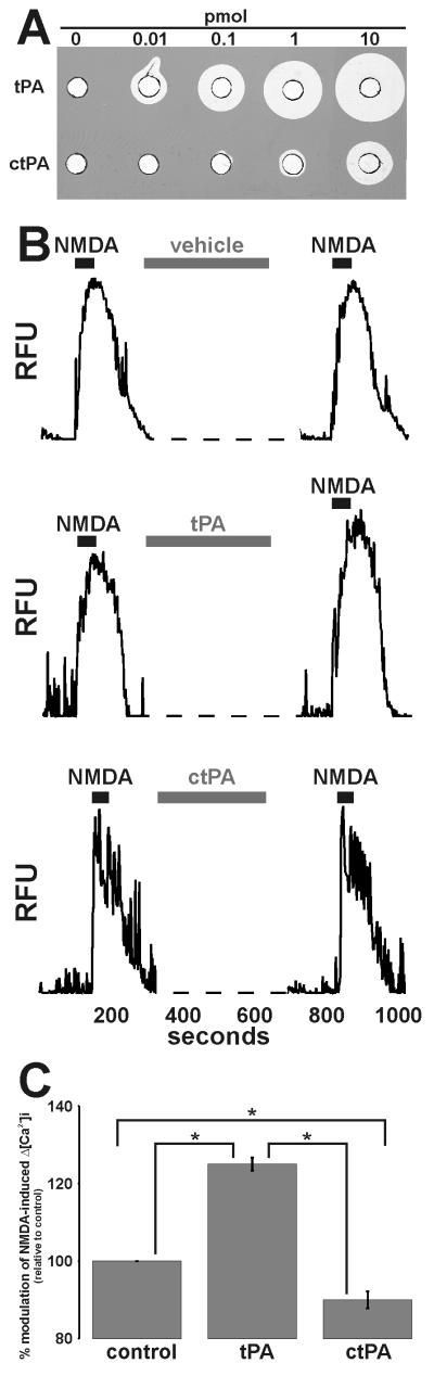
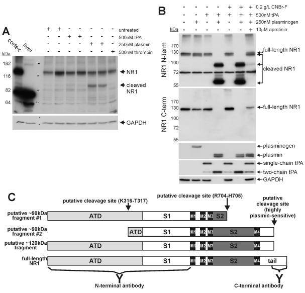
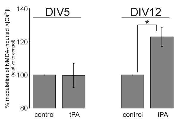
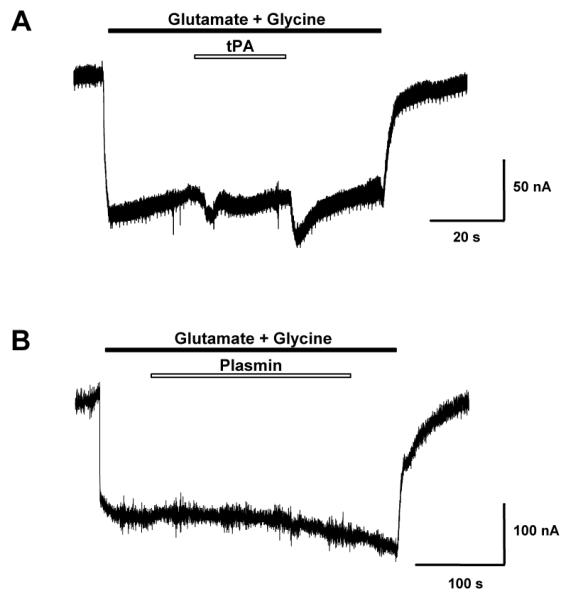
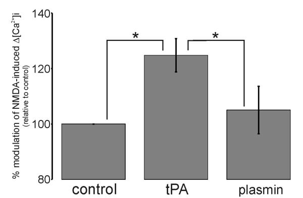
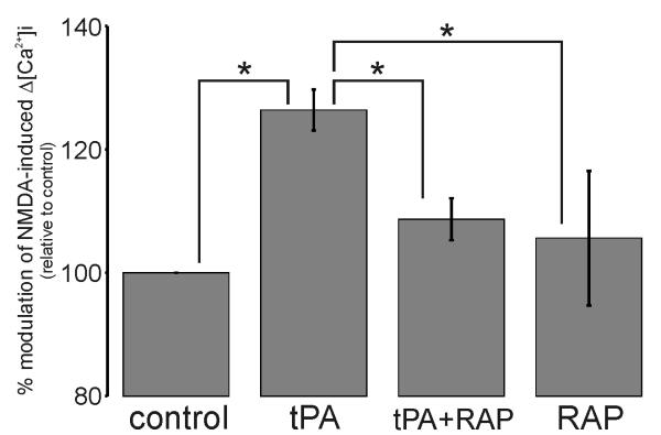
Glutamate is the main excitatory neurotransmitter of the CNS. Tissue-type plasminogen activator (tPA) is recognized as a modulator of glutamatergic neurotransmission. This attribute is exemplified by its ability to potentiate calcium signaling following activation of the glutamate-binding NMDA receptor (NMDAR). It has been hypothesized that tPA can directly cleave the NR1 subunit of the NMDAR and thereby potentiate NMDA-induced calcium influx. In contrast, here we show that this increase in NMDAR signaling requires tPA to be proteolytically active, but does not involve cleavage of the NR1 subunit or plasminogen. Rather, we demonstrate that enhancement of NMDAR function by tPA is mediated by a member of the low-density lipoprotein receptor (LDLR) family. Hence, this study proposes a novel functional relationship between tPA, the NMDAR, a LDLR and an unknown substrate which we suspect to be a serpin. Interestingly, whilst tPA alone failed to cleave NR1, cell-surface NMDARs did serve as an efficient and discrete proteolytic target for plasmin. Hence, plasmin and tPA can affect the NMDAR via distinct avenues. Altogether, we find that plasmin directly proteolyses the NMDAR whilst tPA functions as an indirect modulator of NMDA-induced events via LDLR engagement.


Figures

Figure 1
Figure 1. tPA requires its proteolytic activity to potentiate NMDA-induced Δ[Ca2+]i
Panel A: Increasing amounts of tPA or ctPA were loaded into wells of a fibrin:plasminogen:agarose matrix (Granelli-Piperno & Reich 1978) and incubated at 37°C until proteolyzed zones appeared. Panel B: Representative single cell traces of 25μM NMDA-induced Δ[Ca2+]i before and after 5min perfusion with vehicle, 500nM tPA or 500nM ctPA. Panel C: Collated data demonstrating that 500nM ctPA does not enhance 25μM NMDA-induced Δ[Ca2+]i. Data represents mean ±SEM of the second NMDA-induced Δ[Ca2+]i relative to the first NMDA-induced Δ[Ca2+]i (i.e. % modulation). All data was normalized to control values. The data was pooled from N=61 cells (control), N=84 cells (tPA), N=75 cells (ctPA) over three independent cultures (n=3; * p<0.05).
Figure 2
Figure 2. The NR1 subunit of the NMDAR is a substrate for plasmin, but not tPA
Panel A: Representative cell-based cleavage assay demonstrating that plasmin, but not tPA, can cleave the NR1 subunit of the NMDAR. Cultures were incubated with 500nM tPA, 250nM plasmin or 500nM thrombin at 37°C for 10min. The NR1 content was assessed by immunoblot using the anti-NR1 N-terminal antibody. The membrane was re-probed for GAPDH as a loading control. A total of 6 independent cell-free cleavage assays demonstrated that tPA cannot cleave the NR1 subunit (n=6). Note, the anti-NR1 N-terminal antibody is a “pan” antibody as it recognizes all NR1 variants. Panel B: Representative cell-free cleavage assay demonstrating that plasmin, but not tPA, can cleave the NR1 subunit. tPA−/− mouse cortical lysates were supplemented with CNBr-F, 500nM tPA, plasminogen and aprotinin and incubated at 37°C for 15min. The NR1 content was assessed by immunoblot using both anti-NR1 N-terminal and C-terminal antibodies. To assess the proteolytic actions of tPA and plasmin, membranes were re-probed for plasmin(ogen) and tPA, respectively. The membranes were re-probed for GAPDH as a loading control. Note, the generation of plasmin under cell-free conditions leads to a loss of GAPDH, presumably as a consequence of proteolytic degradation. A total of 7 independent cell-free cleavage assays demonstrate that plasmin, but not tPA, can cleave the NR1 subunit (n=7). Panel C: The NR1 subunit is comprised of an amino-terminal domain (ATD), a bi-lobed ligand binding domain (S1 and S2), four transmembrane segments (M1-M4) and a C-terminal tail. Note, the anti-NR1 N-terminal antibody is raised against the ATD and S1 regions of NR1, whilst the anti-C-terminal antibody is raised against the C-terminal tail. Our cell based cleavage assays indicate that a 90kDa fragment is generated from the naturally presented NR1 molecule, while additional fragments of 120 and 60kDa are generated when cryptic cleavage sites are exposed following cell disruption (i.e. as seen using the cell free cleavage assay). The schematic indicates how these fragments may be generated from the native NR1 molecule. It appears that a highly-plasmin sensitive cleavage site resides near the C-terminus of NR1. Proteolysis at this point yields the 120kDa fragment. The 120kDa fragment is further proteolysed into a 90kDa and a 60kDa fragment that are recognized solely by the N-terminal antibody. Proteolytic generation of the 90kDa fragment is best explained by cleavage at one of two putative cleavage sites: (1) a site within the S2 domain yielding “fragment #1”, or (2) a site within the ATD yielding “fragment #2. The specific cleavage site yielding the 60kDa fragment is unclear and is not shown in the diagram. Several studies have determined the site specificity of plasmin (Harris et al. 2000, Backes et al. 2000, Gosalia et al. 2005, Xue & Seto 2005). This consensus substrate specificity was used to scan the S2 domain for putative plasmin-sensitive cleavage sites. Any putative cleavage sites were then located within the crystal structure of the NR1:NR2A dimeric complex (PDB Identifier # 2A5T) to ensure surface exposure. Via this method, Arginine704-Histidine705 was highlighted as a putative plasmin-sensitive cleavage site with the S2 domain. Using a similar approach, Lysine316-Tyrosine317 was identified as a putative plasmin-sensitive cleavage site with the ATD. In this instance, as the crystal structure of the NR1 ATD has not be resolved, the ATD of the closely related mGluR1 was used (PDB Identifier # 1EWK; (Huggins & Grant 2005)).
Figure 3
Figure 3. tPA potentiation of NMDA-induced Δ[Ca2+]i is a function of in vitro culture age
Collated data demonstrating the potentiating effect of 500nM tPA on 50μM NMDA-induced Δ[Ca2+]i only in mature DIV12 culture. Data represents mean ±SEM of the second NMDA-induced Δ[Ca2+]i relative to the first NMDA-induced Δ[Ca2+]i (i.e. % modulation). All data was normalized to control values. For DIV5 the data was pooled from N=161 cells (control) and N=153 cells (tPA) over four independent cultures (n=4). For DIV12 the data was pooled from N=36 cells (control) and N=56 cells (tPA) over three independent cultures (n=3; * p<0.05). Suppl. Fig.S4 shows the changes in the appearance, NMDA-responsiveness and NMDAR subunit expression that arose during in vitro culture maturation.
Figure 4
Figure 4. tPA does not alter recombinant NMDA-mediated currents in expressed heterologous Xenopus oocytes
Shown is a representative trace of NR1a/2A NMDA-mediated currents expressed in Xenopus oocytes. Cells were voltage clamped at −70mV. NMDA-mediated currents were first evoked with 30μM glutamate and 10μM glycine (black bar), then during NMDAR activation either (A) 1μM tPA or (B) 250nM Plasmin (open white bars) were transiently co-applied. Artifact peaks in Panel A were due to solution exchange. For tPA, a total of 17 independent experiments were conducted. For plasmin, a total of 4 independent experiments were conducted.
Figure 5
Figure 5. tPA potentiates NMDA-induced Δ[Ca2+]i in a plasmin-independent manner
Collated data demonstrating that unlike 500nM tPA, 25nM plasmin fails to potentiate 25μM NMDA-induced Δ[Ca2+]i. Data represents mean ±SEM of the second NMDA-induced Δ[Ca2+]i relative to the first NMDA-induced Δ[Ca2+]i (i.e. % modulation). All data was normalized to control values. The data was pooled from N=134 cells (control), N=64 cells (tPA) and N=66 cells (plasmin) over three independent cultures (n=3; * p<0.05).
Figure 6
Figure 6. Protease Nexin-1 (PN-1) expression increases dramatically during in vitro culture maturation
Cellular protein lysates were obtained from neuronal cultures at different days of in vitro (DIV) maturation. Lysates were collected and analyzed in quadruplicate from two independently-seeded cultures (n=2, N=8). PN-1 expression was assessed by immunoblot analysis. The membrane was re-probed for actin as a loading control. An adult mouse brain cortical lysate was used as a positive control for PN-1 (arrows). Arrowheads indicate probable alternate glycosylated forms of PN-1. Samples were loaded onto the gel in a blinded fashion in the order DIV 7, 5, 9 and 12 as indicated.
Figure 7
Figure 7. A Low-Density Lipoprotein receptor mediates the tPA potentiation of 50μM NMDA-induced Δ[Ca2+]i
Collated data demonstrating potentiation of NMDA-induced Δ[Ca2+]i by 500nM tPA and the ability 500nM RAP to block this effect. Data represents mean ±SEM of the second NMDA-induced Δ[Ca2+]i relative to the first NMDA-induced Δ[Ca2+]i (i.e. % modulation). All data was normalized to control values. The data was pooled from N=83 cells (control), N=78 cells (tPA), N=64 cells (tPA+RAP) and N=55 cells (RAP) over three independent cultures (n=3; * p<0.05).

Similar articles

Cited by

References

    1. Backes BJ, Harris JL, Leonetti F, Craik CS, Ellman JA. Synthesis of positional-scanning libraries of fluorogenic peptide substrates to define the extended substrate specificity of plasmin and thrombin. Nat Biotechnol. 2000;18:187–193. - PubMed
    1. Benchenane K, Castel H, Boulouard M, et al. Anti-NR1 N-terminal-domain vaccination unmasks the crucial action of tPA on NMDA-receptor-mediated toxicity and spatial memory. J Cell Sci. 2007;120:578–585. - PubMed
    1. Boucher P, Gotthardt M, Li WP, Anderson RG, Herz J. LRP: role in vascular wall integrity and protection from atherosclerosis. Science. 2003;300:329–332. - PubMed
    1. Clark RJ, Fischer H, Nevin ST, Adams DJ, Craik DJ. The synthesis, structural characterization, and receptor specificity of the alpha-conotoxin Vc1.1. J Biol Chem. 2006;281:23254–23263. - PubMed
    1. Fernandez-Monreal M, Lopez-Atalaya JP, Benchenane K, et al. Arginine 260 of the amino-terminal domain of NR1 subunit is critical for tissue-type plasminogen activator-mediated enhancement of N-methyl-D-aspartate receptor signaling. J Biol Chem. 2004a;279:50850–50856. - PubMed

Publication types

MeSH terms