Comparative Study
. 2008 Sep 16:8:94.
doi: 10.1186/1471-2229-8-94.

A new picture of cell wall protein dynamics in elongating cells of Arabidopsis thaliana: confirmed actors and newcomers

Affiliations
Comparative Study

A new picture of cell wall protein dynamics in elongating cells of Arabidopsis thaliana: confirmed actors and newcomers

Muhammad Irshad et al. BMC Plant Biol. .

Abstract

Background: Cell elongation in plants requires addition and re-arrangements of cell wall components. Even if some protein families have been shown to play roles in these events, a global picture of proteins present in cell walls of elongating cells is still missing. A proteomic study was performed on etiolated hypocotyls of Arabidopsis used as model of cells undergoing elongation followed by growth arrest within a short time.

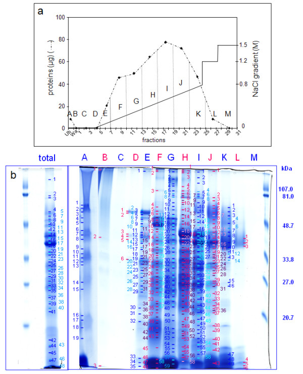
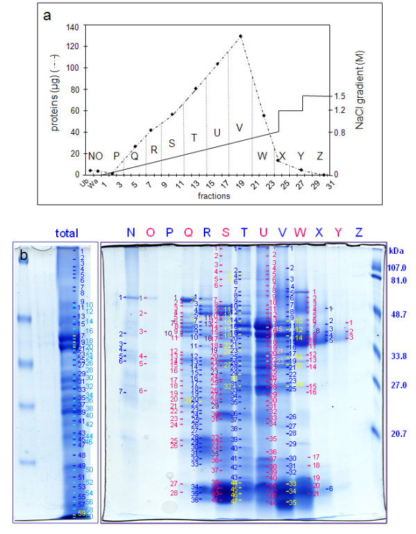
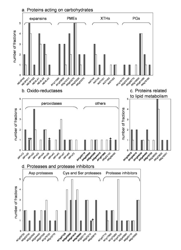
Results: Two developmental stages (active growth and after growth arrest) were compared. A new strategy consisting of high performance cation exchange chromatography and mono-dimensional electrophoresis was established for separation of cell wall proteins. This work allowed identification of 137 predicted secreted proteins, among which 51 had not been identified previously. Apart from expected proteins known to be involved in cell wall extension such as xyloglucan endotransglucosylase-hydrolases, expansins, polygalacturonases, pectin methylesterases and peroxidases, new proteins were identified such as proteases, proteins related to lipid metabolism and proteins of unknown function.

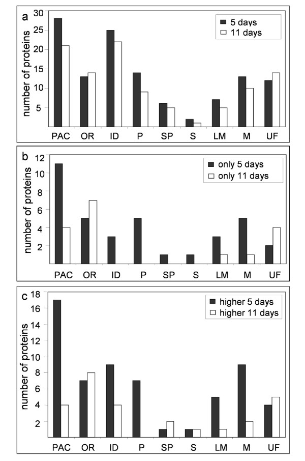
Conclusion: This work highlights the CWP dynamics that takes place between the two developmental stages. The presence of proteins known to be related to cell wall extension after growth arrest showed that these proteins may play other roles in cell walls. Finally, putative regulatory mechanisms of protein biological activity are discussed from this global view of cell wall proteins.


Figures

Figure 1
Figure 1
Analysis of proteins extracted by CaCl2 and LiCl from the cell wall fraction prepared from 5 day-old hypocotyls. a. Separation of proteins by cation exchange chromatography. The graph represents amounts of proteins in each fraction eluted by a NaCl gradient (from 0 M to 0.8 M), followed by two steps at 1.2 M and 1.5 M NaCl. Dotted vertical lines show the grouping of chromatography fractions. Ub stands for unfixed fraction, Wa for washes of the column prior to protein elution, numbering to FPLC fractions, and letters (from A to M) to pools analyzed by 1D-E. b. Separation by 1D-E of the total protein extract (total) and of fractions A to M obtained after cation exchange chromatography. Molecular mass markers are on the right. Numbers refer to bands analyzed by MALDI-TOF MS with successful identification (see Additional data file 1).
Figure 2
Figure 2
Analysis of proteins extracted by CaCl2 and LiCl from the cell wall fraction prepared from 11 day-old hypocotyls.a. Separation of proteins by cation exchange chromatography. The graph shows amounts of proteins in each fraction eluted by a NaCl gradient (from 0 M to 0.8 M) followed by two steps at 1.2 M and 1.5 M NaCl. Dotted vertical lines show the grouping of chromatography fractions. Ub stands for unfixed fraction, Wa for washes of the column prior to protein elution, numbering to FPLC fractions, and letters (from N to Z) to pools analyzed by 1D-E. b. Separation by 1D-E of the total protein extract (total) and of fractions N to Z obtained after cation exchange chromatography. Molecular mass markers are on the right. Numbers refer to bands analyzed by MALDI-TOF MS with successful identification (see Additional data file 2).
Figure 3
Figure 3
Sorting of hypocotyl CWPs in functional classes. Proteins were classified according to their functional domains as described in Experimental procedures: proteins acting on carbohydrates (PAC), oxido-reductases (OR), proteins with interaction domains (ID), proteases (P), structural proteins (SP), proteins involved in signaling (S), proteins related to lipid metabolism (LM), proteins with miscellaneous functions (M), and proteins of yet unknown function (UF). a. Number of proteins found in the nine functional classes in 5 (5 days), and in 11 day-old hypocotyls (11 days). b. Number of proteins found only in 5 (only 5 days), or in 11 day-old (only 11 days) hypocotyls. c. Number of proteins present in higher amount in 5 (higher 5 days) or in 11 day-old (higher 11 days) hypocotyls.
Figure 4
Figure 4
Occurrence of CWPs of selected families of proteins extracted from hypocotyls. a. Families of proteins predicted to encode expansins, PMEs, XTHs and PGs. b. Protein families predicted to encode oxido-reductases. Berberine-bridge enzymes are in bold. c. Protein families predicted to encode proteins related to lipid metabolism. Proteins having a GDSL Lipase/Acylhydrolase domain are in bold. d. Families of proteins predicted to encode proteases or protease inhibitors. Cys proteases are in bold. The number of FPLC fractions in which each protein was identified was counted: black and white bars respectively stand for 5 and 11 day-old etiolated hypocotyls. Stars indicate proteins that have only been identified after separation by 1D-E. Detailed information on the distribution of proteins in the total extract and in FPLC fractions are given in Additional data file 5.

References

    1. Smallwood M, Beven A, Donovan N, Neill S, Peart J, Roberts K, Knox J. Localization of cell wall proteins in relation to the developmental anatomy of the carrot root apex. Plant J. 1994;5:237–246.
    1. Freshour G, Clay RP, Fuller MS, Albersheim P, Darvill AG, Hahn MG. Developmental and tissue-specific structural alterations of the cell-wall polysaccharides of Arabidopsis thaliana roots. Plant Physiol. 1996;110:1413–1429. - PMC - PubMed
    1. Cosgrove DJ. Growth of the plant cell wall. Nat Rev Mol Cell Biol. 2005:850–860. - PubMed
    1. Gendreau E, Traas J, Desnos T, Grandjean O, Caboche M, Hofte H. Cellular basis of hypocotyl growth in Arabidopsis thaliana. Plant Physiol. 1997;114:295–305. - PMC - PubMed
    1. Saibo NJ, Vriezen WH, Beemster GT, Straeten D Van Der. Growth and stomata development of Arabidopsis hypocotyls are controlled by gibberellins and modulated by ethylene and auxins. Plant J. 2003;33:989–1000. - PubMed

Publication types