. 2008 Dec;55(7):1238-50.
doi: 10.1016/j.neuropharm.2008.08.018. Epub 2008 Aug 29.

A NMDA receptor glycine site partial agonist, GLYX-13, simultaneously enhances LTP and reduces LTD at Schaffer collateral-CA1 synapses in hippocampus

Affiliations

A NMDA receptor glycine site partial agonist, GLYX-13, simultaneously enhances LTP and reduces LTD at Schaffer collateral-CA1 synapses in hippocampus

Xiao-lei Zhang et al. Neuropharmacology. 2008 Dec.

Abstract

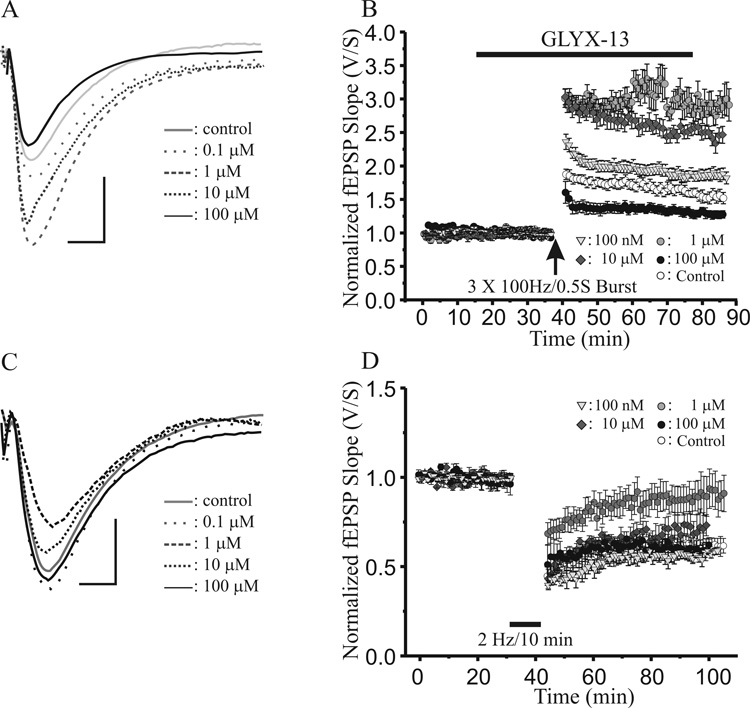
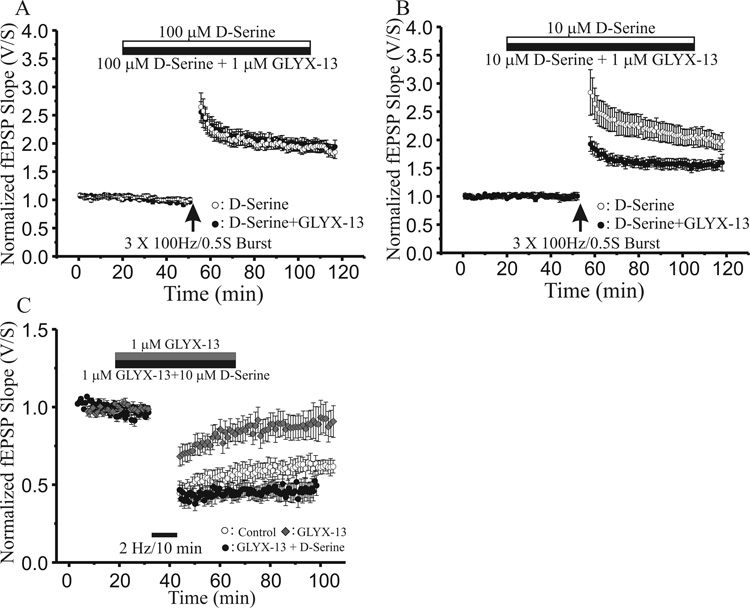
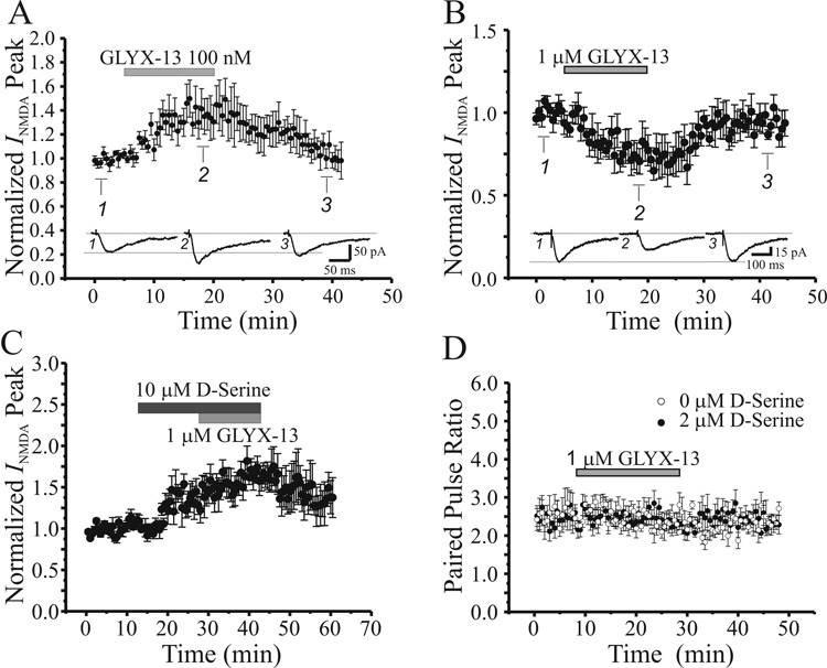
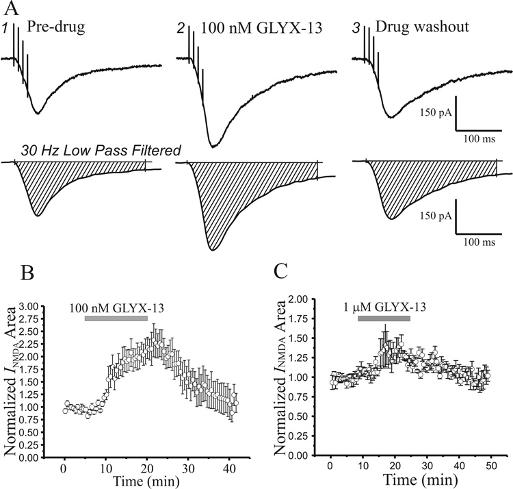
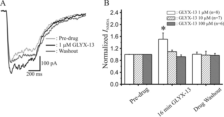
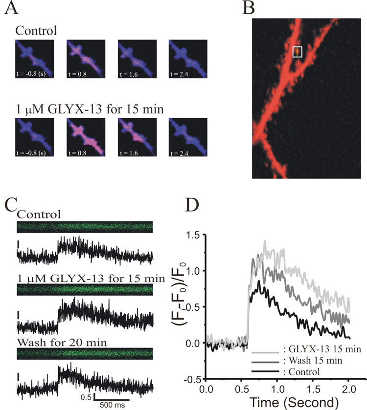
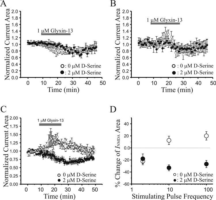
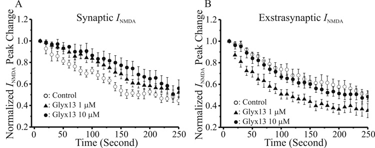
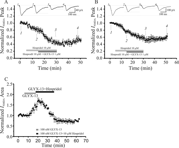
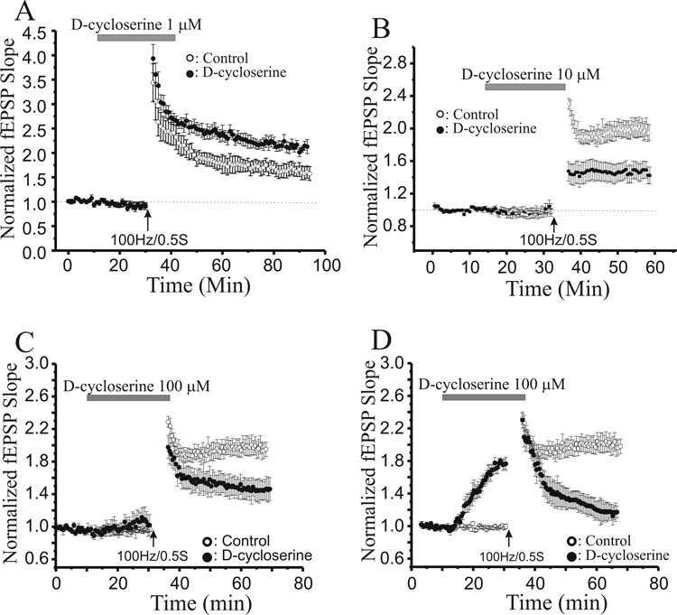
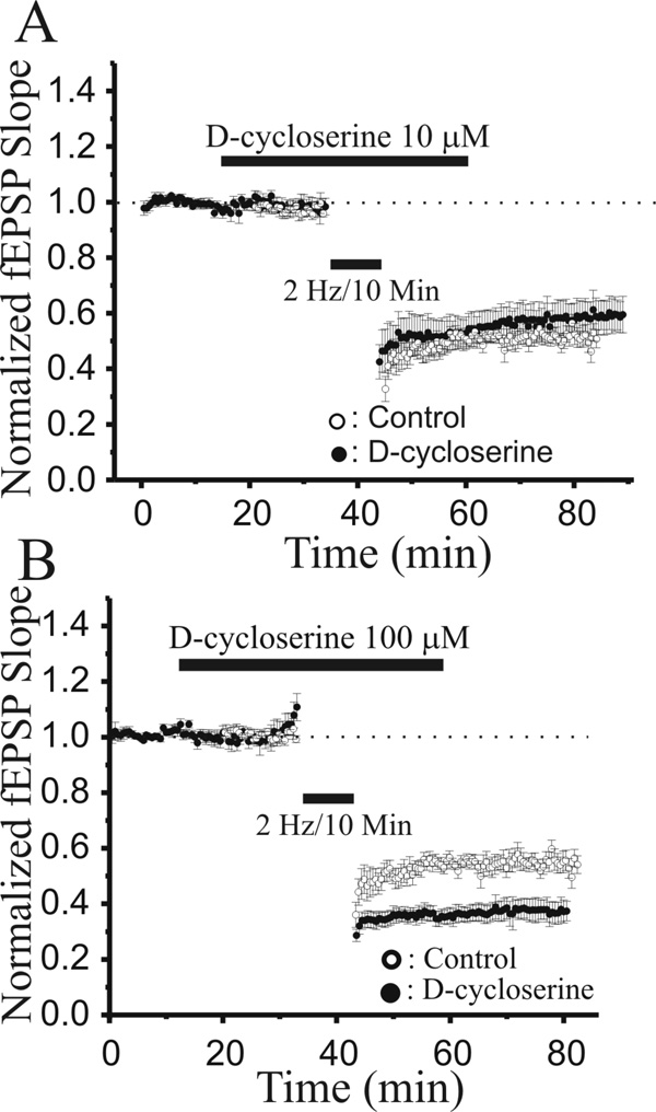
N-methyl-D-aspartate glutamate receptors (NMDARs) are a key route for Ca2+ influx into neurons important to both activity-dependent synaptic plasticity and, when uncontrolled, triggering events that cause neuronal degeneration and death. Among regulatory binding sites on the NMDAR complex is a glycine binding site, distinct from the glutamate binding site, which must be co-activated for NMDAR channel opening. We developed a novel glycine site partial agonist, GLYX-13, which is both nootropic and neuroprotective in vivo. Here, we assessed the effects of GLYX-13 on long-term synaptic plasticity and NMDAR transmission at Schaffer collateral-CA1 synapses in hippocampal slices in vitro. GLYX-13 simultaneously enhanced the magnitude of long-term potentiation (LTP) of synaptic transmission, while reducing long-term depression (LTD). GLYX-13 reduced NMDA receptor-mediated synaptic currents in CA1 pyramidal neurons evoked by low frequency Schaffer collateral stimulation, but enhanced NMDAR currents during high frequency bursts of activity, and these actions were occluded by a saturating concentration of the glycine site agonist d-serine. Direct two-photon imaging of Schaffer collateral burst-evoked increases in [Ca2+] in individual dendritic spines revealed that GLYX-13 selectively enhanced burst-induced NMDAR-dependent spine Ca2+ influx. Examining the rate of MK-801 block of synaptic versus extrasynaptic NMDAR-gated channels revealed that GLYX-13 selectively enhanced activation of burst-driven extrasynaptic NMDARs, with an action that was blocked by the NR2B-selective NMDAR antagonist ifenprodil. Our data suggest that GLYX-13 may have unique therapeutic potential as a learning and memory enhancer because of its ability to simultaneously enhance LTP and suppress LTD.


Figures

Fig. 1
Fig. 1
0.1–10 µM GLYX-13 enhances the magnitude of long-term potentiation (LTP), while 1–10 µM GLYX-13 reduces long-term depression (LTD) of synaptic transmission at Schaffer collateral-CA1 synapses. A: Representative excitatory postsynaptic potentials (EPSP) after induction of control LTP (solid grey), compared to LTP in GLYX-13 at the indicated concentrations. (Calibration bar for A and C 0.5 mV/10 msec) B: Time course of experiments comparing LTP induced by a high frequency stimulus train (3×100Hz/500ms; arrow) at Schaffer collateral-CA1 synapses in control, untreated slices (open circles; n=15), compared to slices pretreated with 100 nM (triangle; n=8), 1 µM (grey circles; n=8), 10 µM (diamonds; n=6), or 100 µM (filled circles; n=8) GLYX-13 (solid bar). (Each point mean ± SEM of normalized field e.p.s.p. slope of n slices.) C: Representative excitatory postsynaptic potentials (EPSP) after induction of control LTP (solid grey), compared to LTP in GLYX-13 at the indicated concentrations. D: Time course of LTD induced by a low frequency stimulus train (2Hz/10min; solid bar) at Schaffer collateral-CA1 synapses in slices pre-treated with 100 nM (triangles; n=8), 1 µM (grey circles; n=7), 10µM (diamonds; n=6), or 100 µM (filled circles; n=6) GLYX-13 (solid bar), compared to control, untreated slices (open circles; n=16). (Each point mean ± SEM of normalized field e.p.s.p. slope of n slices.)
Fig. 2
Fig. 2
The NMDAR glycine site full agonist D-serine occludes the effects of GLYX-13 on both LTP and LTD. A: Time course of experiments comparing LTP induced by a high frequency stimulus train (3×100Hz/500ms; arrow) at Schaffer collateral-CA1 synapses in slices in presence of 100 µM D-serine alone (open circles; n=7), versus slices where 100 µM D-serine was coapplied with 1 µM GLYX-13 (filled circles; n=7). (Each point mean ± SEM of normalized field e.p.s.p. slope of n slices.) B: Time course of experiments comparing LTP induced by a high frequency stimulus train (3×100Hz/500ms; arrow) at Schaffer collateral-CA1 synapses in slices in presence of 10 µM D-serine alone (open circles; n=8), versus slices where 10 µM D-serine was co-applied with 1 µM GLYX-13 (filled circles; n=8). C: Time course of experiments comparing LTD induced by a low frequency stimulus train (2Hz/10min; solid bar) at Schaffer collateral-CA1 synapses in control slices (open circles; n=6), compared to slices bathed in 1 µM GLYX-13 alone (diamonds; n=7) or 1 µM GLYX-13 plus 10 µM D-serine (filled circles; n=7).
Fig. 3
Fig. 3
A low concentration of GLYX-13 enhances, and a 10-fold higher concentration reduces, postsynaptic NMDA receptor-mediated Schaffer collateral-evoked excitatory postsynaptic currents (EPSCs) by binding a D-serine sensitive site, without altering presynaptic transmission. A: Time course of the effect of GLYX-13 (100 nM; grey bar) on the NMDA component of Schaffer collateral-evoked EPSCs in CA1 pyramidal neurons (n=8). Each point is the mean ± SEM of EPSC peak amplitude of n cells. Insets: Signal-averaged sample EPSCs recorded at the times indicated. B: Time course of the effect of GLYX-13 (1 µM; grey bar) on the NMDA component of Schaffer collateral-evoked EPSCs in CA1 pyramidal neurons (n=8). Each point is the mean ± SEM of EPSC peak amplitude of n cells. Insets: Signal-averaged sample EPSCs recorded at the times indicated. C: Time course of the effect of GLYX-13 (1 µM; light grey bar) in the presence of D-serine (10 µM; dark grey bar) on the NMDA component of Schaffer collateral-evoked EPSCs in CA1 pyramidal neurons (n=8). Each point is the mean ± SEM of EPSC peak amplitude of n cells. D: Time course of the lack of effect of GLYX-13 (1 µM; grey bar) on paired-pulse ratio in normal ACSF (open circles; n=8) and in the presence of D-serine (2 µM; filled circles; n=7). Each point is the mean ± SEM of EPSC peak amplitude of n cells.
Fig. 4
Fig. 4
GLYX-13 produces an enhancement of NMDA receptor-mediated summed EPSCs evoked by high-frequency burst stimulation. A: Sample EPSCs (top row) before, during application and 30 min after washout of GLYX-13 (100 nM) evoked by a burst of 4 Schaffer collateral stimuli given at a frequency of 100 Hz, and low-pass (30Hz) filtered EPSCs to eliminate stimulus artifacts for measurement (bottom row) of total NMDA current from CA1 pyramidal neurons in slices bathed in CNQX (20 µM) plus bicuculline (10 µM) in Mg2+-free ACSF. B: Time course of the effect of GLYX-13 (100 nM; grey bar) on total burst-induced NMDA EPSC area in normal ACSF (n=8). Each point is the mean ± SEM of EPSC peak amplitude normalized to starting amplitude in n cells. C: Time course of the effect of GLYX-13 (1 µM; grey bar; n=8) on burst-induced NMDA EPSC area in normal ACSF. Each point is the mean ± SEM of EPSC peak amplitude normalized to starting amplitude in n cells.
Fig. 5
Fig. 5
GLYX-13 also enhances NMDA receptor-gated conductances during longer high-frequency stimulus bursts of the type that elicits LTP. A: Sample low-pass (30Hz) filtered NMDAR-mediated EPSCs evoked by 100Hz/0.5sec bursts of Schaffer collateral stimulation in CA1 pyramidal neurons before (Ctl), during 16 min application of GLYX-13 (1 µM) and 30 min after washout. B: Mean ± SEM normalized current area of NMDAR EPSCs evoked by 50Hz/0.5sec Schaffer collateral stimulus trains before (Pre-drug), after 16 min application of GLYX-13 (16 min GLYX-13), and 30 min after drug washout, where 1 µM GLYX-13 significantly enhanced NMDAR EPSCs evoked by prolonged high-frequency trains of stimuli (*, P<0.05, paired t-test, n=# slices).
Fig. 6
Fig. 6
GLYX-13 enhances burst-induced NMDA receptor-dependent Ca2+ influx into individual dendritic spines of CA1 pyramidal neurons. A: Sample two-photon image of the increase in [Ca2+] in dendritic spines of a typical CA1 pyramidal neuron, measured with the indicator Calcium Green, in normal ACSF (top row) versus in GLYX-13 (1 µM; bottom row). B: Sample two-photon image of a CA1 pyramidal neuron dendrite using Alexafluor 594. The cross-section scanned in panel c is indicated in the box. C: Two-photon line scans of [Ca2+] in the single dendritic spine shown in b, in normal ACSF (Control), after 15 min in GLYX-13 (1 µM GLYX-13 for 15’), and 20 min after drug washout (Wash for 20’). D: Signal-averages of 5–7 line scans from a single dendritic spine of a CA1 pyramidal neuron in normal ACSF (Control), after 15 min in GLYX-13 (1 µM; GLYX-13 15’), and after 15 min washout (Wash for 15’).
Fig. 7
Fig. 7
GLYX-13 selectively enhances NMDA receptor-gated conductances during later pulses in a short high-frequency stimulus burst. A: Time course of the effect of GLYX-13 (1 µM; grey bar) on the first pulse of a 4 stimulus, 2 Hz train, in normal ACSF (open circles; n=8) and in presence of D-serine (2 µM; filled circles; n=8). B: Time course of the effect of GLYX-13 (1 µM; grey bar) on the fourth pulse of a 4 stimulus, 2 Hz train, in normal ACSF (open circles; n=8) and in presence of D-serine (2 µM; filled circles; n=9). C: Time course of the effect of GLYX-13 (1 µM; grey bar) on the NMDA EPSC area evoked by a 4 stimulus, 100 Hz burst, in normal ACSF (open circles; n=8) and in presence of D-serine (2 µM; filled circles; n=8). D: Mean ± SEM percent change in the NMDA EPSC elicited by GLYX-13 (1 µM) as a function of Schaffer collateral stimulus frequency, in normal ACSF (open circles; n=8) and in D-serine (2 µM; filled circles; n=8).
Fig. 8
Fig. 8
GLYX-13 reduces openings of synaptic NMDA receptors, while increasing openings of extrasynaptic NMDA receptors. A: Time course of the blockade of NMDA receptor-dependent single shock-evoked synaptic EPSCs by the open channel blocker MK-801 (4 µM) in control slices (open circles), versus slices in the presence of 1 µM (filled triangles) or 10 µM (filled circles) GLYX-13. Each point is the mean ± SEM of EPSC peak amplitude normalized to starting amplitude in n cells. B: Time course of the blockade of burst-evoked (4 pulses/100Hz) extrasynaptic NMDA receptor-dependent EPSCs by MK-801 (4 µM), elicited once synaptic NMDA channel block had plateaued, in control slices (open circles), versus slices in the presence of 1 µM (filled triangles) or 10 µM (filled circles) GLYX-13. Each point is the mean ± SEM of EPSC peak amplitude normalized to starting amplitude in n cells.
Fig. 9
Fig. 9
GLYX-13 enhances NMDA receptor-gated extrasynaptic conductances by acting on NR2B-containing NMDA receptors, while its suppression of synaptic NMDA currents is NR2Bindependent. A: Time course of the effect of GLYX-13 (1 µM; dark bar) on NMDA receptor-dependent single shock-evoked EPSCs in the presence of the NR2B-selective NMDA receptor antagonist ifenprodil (10 µM; light bar). The suppression of single-shock evoked NMDA currents by GLYX-13 is not affected by blockade of NR2B-containing NMDA receptors. (Insets are single shock-evoked NMDAR EPSCs at the indicated times) B: Time course of the effect of GLYX-13 (1 µM; dark bar) on NMDA receptor-dependent burst-evoked EPSCs in the presence of the NR2B-selective NMDA receptor antagonist ifenprodil (10 µM; light bar). The enhancement of burst-evoked NMDA EPSCs normal elicited by GLYX-13 is converted to a depression by blockade of NR2B-containiong NMDA receptors. (Insets are burst-evoked NMDAR EPSCs at the indicated times)
Fig. 10
Fig. 10
A low concentration of D-cycloserine (DCS, 1µM) enhances the magnitude of LTP, while a ten and hundred-fold higher concentrations of reduce LTP. A: Time course of the effect of 1 µM DCS on LTP induced by high frequency Schaffer collateral stimulus trains (3×100Hz/200ms; arrow) in slices pre-treated with DCS (1 µM; grey bar; filled circles; n=10), compared to control, untreated slices (open circles; n=8). B: Time course of the effect of 10 µM DCS on LTP induced by high frequency Schaffer collateral stimulus trains (3×100Hz/500ms; arrow) in slices pre-treated with DCS (10 µM; grey bar; filled circles; n=9), compared to control, untreated slices (open circles; n=8). C: Time course of the effect of 100 µM DCS on LTP induced by high frequency Schaffer collateral stimulus trains (3×100Hz/500ms; arrow) in slices where pre-treatment with DCS (100 µM; grey bar; filled circles; n=4) produced no effect alone, compared to control, untreated slices (open circles; n=8). D: Time course of the effect of 100 µM DCS on LTP induced by high frequency Schaffer collateral stimulus trains (3×100Hz/500ms; arrow) in slices where pre-treatment with DCS (100 µM; grey bar; filled circles; n=5) produced marked potentiation on its own, compared to control, untreated slices (open circles; n=8). (Each point is the mean ± SEM of normalized field e.p.s.p. slope of n slices.)
Fig. 11
Fig. 11
10 µM DCS did not alter the magnitude of LTD, while 100µM DCS enhanced LTD. A: Time course of the effect of 100 µM DCS on LTD induced by a low frequency stimulus train (2Hz/10min; solid bar) at Schaffer collateral-CA1 synapses in slices pre-treated with DCS (10 µM; grey bar; filled circles; n=10), compared to control, untreated slices (open circles; n=7). B: Time course of the effect of 100 µM DCS on LTD induced by a low frequency stimulus train (2Hz/10min; solid bar) at Schaffer collateral-CA1 synapses in slices pre-treated with DCS (100 µM; filled circles; n=8), compared to control, untreated slices (open circles; n=7). (Each point is the mean ± SEM of normalized field e.p.s.p. slope of n slices.)

References

    1. Akhondzadeh S. Hippocampal synaptic plasticity and cognition. J. Clin. Pharm. Ther. 1999;24:241–248. - PubMed
    1. Asztely F, Erdemli G, Kullmann DM. Extrasynaptic glutamate spillover in the hippocampus: dependence on temperature and the role of active glutamate uptake. Neuron. 1997;18:281–293. - PubMed
    1. Billard JM, Rouaud E. Deficit of NMDA receptor activation in CA1 hippocampal area of aged rats is rescued by D-cycloserine. Eur. J. Neurosci. 2007;25:2260–2268. - PubMed
    1. Brickley SG, Misra C, Mok MH, Mishina M, Cull-Candy SG. NR2B and NR2D subunits coassemble in cerebellar Golgi cells to form a distinct NMDA receptor subtype restricted to extrasynaptic sites. J. Neurosci. 2003;23:4958–4966. - PMC - PubMed
    1. Collingridge GL, Kehl SJ, McLennan H. Excitatory amino acids in synaptic transmission in the Schaffer collateral-commissural pathway of the rat hippocampus. J. Physiol. 1983;334:33–46. - PMC - PubMed

Publication types

MeSH terms