. 2008 Nov 14;283(46):31813-22.
doi: 10.1074/jbc.M805935200. Epub 2008 Sep 16.

Differential roles of the COOH termini of AAA subunits of PA700 (19 S regulator) in asymmetric assembly and activation of the 26 S proteasome

Affiliations

Differential roles of the COOH termini of AAA subunits of PA700 (19 S regulator) in asymmetric assembly and activation of the 26 S proteasome

Thomas G Gillette et al. J Biol Chem. .

Abstract

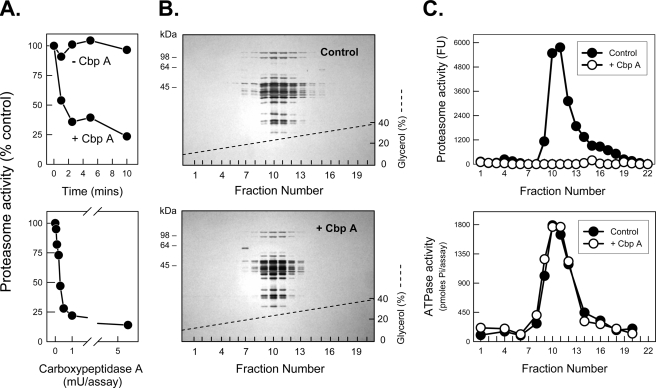
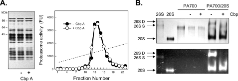
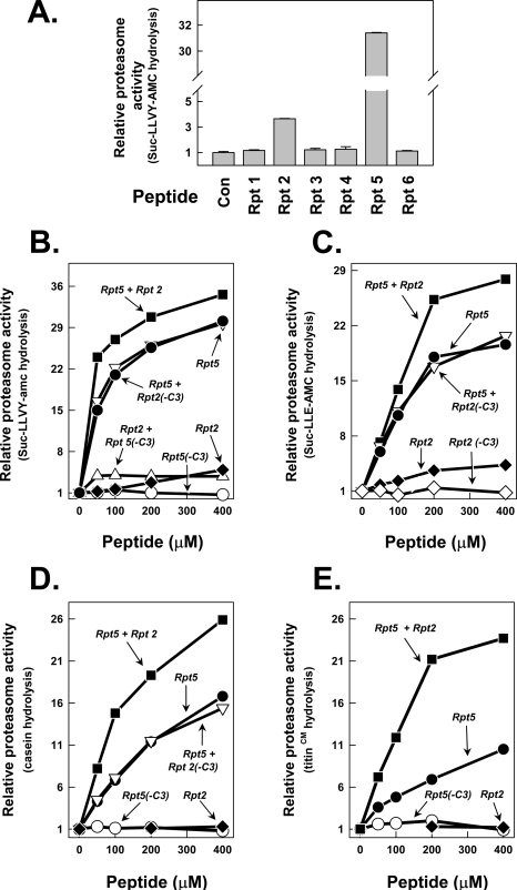
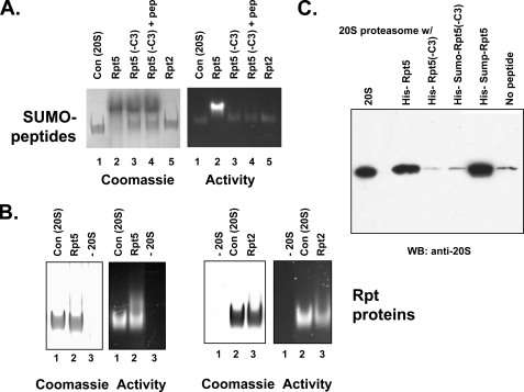
The 26 S proteasome is an energy-dependent protease that degrades proteins modified with polyubiquitin chains. It is assembled from two multi-protein subcomplexes: a protease (20 S proteasome) and an ATPase regulatory complex (PA700 or 19 S regulatory particle) that contains six different AAA family subunits (Rpt1 to -6). Here we show that binding of PA700 to the 20 S proteasome is mediated by the COOH termini of two (Rpt2 and Rpt5) of the six Rpt subunits that constitute the interaction surface between the subcomplexes. COOH-terminal peptides of either Rpt2 or Rpt5 bind to the 20 S proteasome and activate hydrolysis of short peptide substrates. Simultaneous binding of both COOH-terminal peptides had additive effects on peptide substrate hydrolysis, suggesting that they bind to distinct sites on the proteasome. In contrast, only the Rpt5 peptide activated hydrolysis of protein substrates. Nevertheless, the COOH-terminal peptide of Rpt2 greatly enhanced this effect, suggesting that proteasome activation is a multistate process. Rpt2 and Rpt5 COOH-terminal peptides cross-linked to different but specific subunits of the 20 S proteasome. These results reveal critical roles of COOH termini of Rpt subunits of PA700 in the assembly and activation of eukaryotic 26 S proteasome. Moreover, they support a model in which Rpt subunits bind to dedicated sites on the proteasome and play specific, nonequivalent roles in the asymmetric assembly and activation of the 26 S proteasome.


Figures

FIGURE 1.
FIGURE 1.
Carboxypeptidase A inhibits PA700 stimulation of 20 S proteasome activity. A (top), PA700 (5 pmol) was incubated at 25 °C in the presence and absence of 13 milliunits of pancreatic CbpA. At the indicated times, PA700 was assayed for ATP-dependent stimulation of 20 S proteasome-catalyzed hydrolysis of Suc-Leu-Leu-Val-Tyr-AMC. Proteasome activity stimulated by untreated PA700 was assigned a value of 100% (control), and other activities are expressed as a percentage of that value. Untreated PA700 stimulated proteasome activity by 44-fold in this experiment. A (bottom), PA700 (5 pmol) was incubated with the indicated concentrations of CbpA for 5 min at 25 °C and then assayed for proteasome stimulatory activity as above. Untreated PA700 stimulated proteasome activity by 49-fold in this experiment. B and C, PA700 was incubated for 10 min with and without 10 milliunits of CbpA, as indicated. Samples were then subjected to 10–40% glycerol density gradient centrifugation, as described previously (32). Fractions were either subjected to SDS-PAGE and stained with Coomassie Blue (B) or assayed for proteasome stimulatory activity, expressed as fluorescent units (Fu), and ATPase activity (C). The dashed line denotes the glycerol concentration gradient from 10 to 40%.
FIGURE 2.
FIGURE 2.
Carboxypeptidase A inhibits assembly and activation of the 26 S proteasome. A (left), SDS-PAGE of purified 26 S proteasome without incubation or after a 10-min incubation with (+) and without (-) CbpA. A (right), 26 S proteasome was incubated in the presence and absence of CbpA and subjected to glycerol density gradient centrifugation. Fractions were assayed for proteasome activity by hydrolysis of Suc-Leu-Leu-Val-Tyr-AMC and expressed as fluorescent units (Fu). B, purified PA700 was preincubated with (+, lanes 5 and 7) and without (-, lanes 4 and 6) CbpA and then incubated with purified 20 S proteasome in the presence of ATP. After 30 min, samples were subjected to native PAGE, as described previously (32). Gels were stained with Coomassie Blue (top) or overlaid with Suc-Leu-Leu-Val-Tyr-AMC, incubated for 10 min, and exposed to UV light (bottom). Purified 26 S proteasome (lane 1), 20 S proteasome (lane 2), and PA700 were electrophoresed as standards. 26S-D, doubly capped 26 S proteasome; 26S-S, singly capped 26 S proteasome.
FIGURE 3.
FIGURE 3.
COOH-terminal peptides of Rpt2 and Rpt5 selectively stimulate 20 S proteasome activity. Peptides corresponding to the COOH-terminal residues of AAA subunits of PA700 (Rpt1 to -6) were synthesized and purified as described under “Experimental Procedures.” A, indicated peptides (400 μm) were incubated with 20 nm purified bovine 20 S proteasome; proteasome activity against Suc-Leu-Leu-Val-Tyr-AMC was measured as described under “Experimental Procedures.” Proteasome activity in the absence of peptide was set at a relative value of 1.0, and other activities are expressed as a -fold increase of that value. B and C, individual peptides at the indicated concentrations were incubated with 20 nm 20 S proteasome for assay of proteasome activation as in A. Where a second peptide is indicated, the concentration of the second peptide is 400 μm. B, Suc-Leu-Leu-Val-Tyr-AMC substrate; C, Suc-Leu-Leu-Glu-AMC substrate. D and E, Rpt peptides, as described for B and C, were assayed for stimulation of 20 S proteasome against casein (D) and carboxymethylated (CM) titin (E). All values are means of triplicate assays. Similar results were obtained in at least three independent experiments.
FIGURE 4.
FIGURE 4.
Binding of COOH-terminal Rpt peptides to the 20 S proteasome. A and B, purified 20 S proteasome and recombinant proteins were incubated at 37 °C for 15 min and then subjected to native PAGE. A, SUMO-Rpt COOH-terminal peptide fusion proteins, produced as described under “Experimental Procedures,” were incubated at 300 μm with 650 nm 20 S proteasome. Lane 1, 20 S proteasome alone; lane 2, 20 S proteasome and SUMO-Rpt5; lane 3, 20 S proteasome and SUMO-Rpt5(-C3); lane 4, 20 S proteasome and SUMO-Rpt5(-C3) and Rpt5 peptide (100 μm); lane 5, 20 S proteasome and SUMO-Rpt2. The gel was overlaid with Suc-Leu-Leu-Val-Tyr-AMC for identification of proteasome activity (right) and stained with Coomassie Blue (left). B, recombinant Rpt proteins, produced as described under “Experimental Procedures,” were incubated at 5 μm with 450 nm 20 S proteasome and analyzed as in A. Left pair of panels, Rpt5(lanes 1–3). Lane 1, 20 S proteasome alone; lane 2, 20 S proteasome and Rpt5; lane 3, Rpt5 alone. Right pair of panels, Rpt2 (lanes 1–3). Lane 1, Rpt2 alone; lane 2, 20 S proteasome alone; lane 3, 20 S proteasome and Rpt2. Gels were either assayed for proteasome activity or stained with Coomassie Brilliant Blue as above. C, purified 20 S proteasome (285 nm) was incubated with indicated His-tagged recombinant proteins (11 μm for Rpt5 and Rpt5-C3 and 90 μm for SUMO-Rpt5 and SUMO-Rpt5-C3) and subjected to pull-down on nickel beads as described under “Experimental Procedures.” Bound and eluted material was Western blotted for 20 S proteasome. Lane 1 (20 S) shows 5% of the input 20 S proteasome; the remaining lanes show 20 S proteasome pulled down by the indicated samples.
FIGURE 5.
FIGURE 5.
Dopa-Rpt activating peptides interact with specific α subunits of the 20 S proteasome. A, purified 20 S proteasome (200 nm) was incubated at the indicated concentrations with biotinylated Dopa-containing Rpt5 and Rpt5(-C3) peptides. Cross-linking was performed as described under “Experimental Procedures.” Lane 1, a control reaction in which cross-linking was not induced with oxidant. Western blot analysis (WB) after SDS-PAGE shows the cross-linked product detected by Neutravidin-HRP (top) and a loading control for 20 S proteasome β5 subunit (bottom). B, samples of cross-linking reactions, performed as in A, were applied to monomeric avidin beads. S, starting cross-linked material; U, unbound fraction; B, bound fraction. In each case, 20% of total fraction was applied to the gel. Western blot analysis after SDS-PAGE shows the cross-linked product detected by neutravidin-HRP. C, two-dimensional PAGE analysis of enriched Dopa peptide-20 S proteasome cross-linked products. Western blotting of 20 S proteasome control (top) and Dopa peptide-20 S proteasome cross-link for the indicated Rpt peptides and proteasome subunits using subunit-specific antibodies (middle). Western blotting for the indicated cross-linked proteasome subunit using neutravidin-HRP (bottom).
FIGURE 6.
FIGURE 6.
Model for PA700 binding to and activation of the proteasome via COOH termini of Rpt2 and Rpt5 subunits. ATP binding to Rpt subunits of PA700 promotes conformational changes that optimize registration of COOH termini of Rpt2 and Rpt5 for interaction with cognate binding sites at proteasome subunits α7 and α4, respectively. Binding of these subunits induces conformational changes in α subunits of proteasome, resulting in gate opening and an increase in substrate access to catalytic sites. Peptides corresponding to the COOH termini of Rpt2 and/or Rpt5 can bind directly to their respective sites on α subunits of the proteasome to promote gate opening and proteasome activation by the same mechanism. Because no conformational change of PA700 is required for peptide binding, proteasome activation is ATP-independent. The nomenclature for 20 S proteasome subunits corresponds to that of Groll et al. (9).

References

    1. Coux, O., Tanaka, K., and Goldberg, A. L. (1996) Annu. Rev. Biochem. 65 801-847 - PubMed
    1. Voges, D., Zwickl, P., and Baumeister, W. (1999) Annu. Rev. Biochem. 68 1015-1068 - PubMed
    1. Pickart, C. M., and Cohen, R. E. (2004) Nat. Rev. Mol. Cell Biol. 5 177-187 - PubMed
    1. Gillette, T. G., and DeMartino, G. N. (2007) Cell 129 659-662 - PubMed
    1. DeMartino, G. N., and Slaughter, C. A. (1999) J. Biol. Chem. 274 22123-22126 - PubMed

Publication types

LinkOut - more resources