. 2008 Sep 23;105(38):14567-72.
doi: 10.1073/pnas.0800589105. Epub 2008 Sep 16.

Monkeypox virus evades antiviral CD4+ and CD8+ T cell responses by suppressing cognate T cell activation

Affiliations

Monkeypox virus evades antiviral CD4+ and CD8+ T cell responses by suppressing cognate T cell activation

Erika Hammarlund et al. Proc Natl Acad Sci U S A. .

Abstract

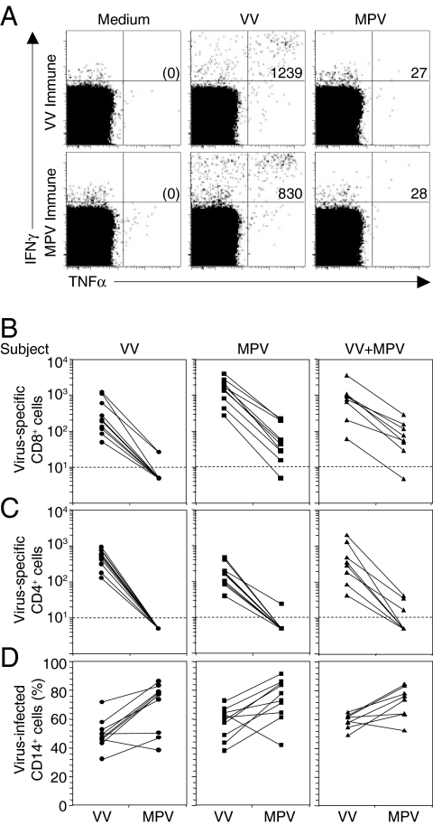
Monkeypox virus (MPV) is a virulent human pathogen that has gained increased attention because of its potential use as a bioterrorism agent and inadvertent introduction into North America in 2003. The US outbreak also provided an important opportunity to study MPV-specific T cell immunity. Although MPV-specific CD4(+) and CD8(+) T cells could recognize vaccinia virus (VV)-infected monocytes and produce inflammatory cytokines such as IFNgamma and TNFalpha, they were largely incapable of responding to autologous MPV-infected cells. Further analysis revealed that, unlike cowpox virus (CPV), MPV did not interfere with MHC expression or intracellular transport of MHC molecules. Instead, MPV-infected cells were capable of preventing T cell receptor (TcR)-mediated T cell activation in trans. The ability to trigger a state of nonresponsiveness represents a unique MHC-independent mechanism for blocking antiviral T cell activation and inflammatory cytokine production and is likely an important attribute involved with viral dissemination in the infected host.


Conflict of interest statement

The authors declare no conflict of interest.

Figures

Fig. 1.
Fig. 1.
MPV immune evasion from orthopoxvirus-specific T cells. (A) Antiviral CD8+ T cell responses from a VV-immune (6 months post-VV infection) or a MPV-immune (4 months post-MPV infection) subject were measured by ICCS after 18 h of stimulation with VV or MPV (MOI of 0.3) with Brefeldin A added for the last 6 h of stimulation. PBMCs were gated on CD8β+CD4 T cells. The numbers in the upper right quadrants depict the frequency of virus-specific IFNγ+TNFα+ T cells per million CD8+ T cells identified after background subtraction from control wells containing Medium alone (shown in parenthesis). (B) Virus-specific CD8+ T cell responses against VV or MPV were determined as in A, using PBMCs from VV-immune subjects (VV; 4–6 months postinfection, n = 10), MPV-immune subjects (MPV; 3–31 months post-MPV infection, n = 10) or VV-immune subjects who contracted MPV infection (VV+MPV; 3–13 months post-MPV infection, n = 8). (C) Virus-specific CD4+CD8β T cell responses against VV or MPV were determined as in A. (D) Monocytes were identified based on forward and side scatter characteristics and CD14 surface expression. The percentage of CD14+ monocytes infected with VV or MPV was determined after 18 h of infection, using a polyclonal anti-orthopoxvirus antibody (40).
Fig. 2.
Fig. 2.
Inhibition of antiviral T cell responses by MPV is not mediated by MHC down-regulation. (A) After 16 h of infection, virus-infected CD14+ cells were identified by intracellular staining for OPV antigens as described in ref. . Surface expression of MHC class I (HLA-A, -B, and -C) on virus-infected primary human monocytes was down-regulated by CPV, but not by VV or MPV. Surface expression of MHC class II (HLA-DR) was unaltered by infection with CPV, VV, or MPV. (B) MHC class I assembly and transport was measured in OPV-infected cells. HeLa cells were uninfected (uninfected* indicates a lighter scanned image of the same experiment) or infected with VV, MPV, or CPV for 5 h (starved for the last 1 h) and pulse-labeled for 20 min, and the labels were chased for 0, 30, or 60 min as indicated. More than 90% of HeLa cells were infected (data not shown), and cell lysates were immunoprecipitated with anti-MHC class I antibody, W6/32. The precipitated material was treated with EndoH and separated by SDS/PAGE. HC, heavy chain; ER, EndoH-resistant band; ES, EndoH-sensitive band. These data show one of four independent experiments, each with comparable results. (C) Stability of MHC class I heavy chain was determined in OPV-infected cells. HeLa cells were infected with VV or MPV for 5 h; starved for the last 1 h in the presence or absence of proteasomal inhibitor, MG132; and pulse-labeled for 20 min, and the labels were chased for 0, 30, or 60 min in the continued presence or absence of MG132. The lysates were then immunoprecipitated with polyclonal anti-MHC class I antibody, K455, followed by EndoH treatment and SDS/PAGE. The open arrowheads indicate the presence of nonspecific low molecular weight proteins that were immunoprecipitated with either anti-MHC class I or anti-CD44 antibodies (see Fig. S1).
Fig. 3.
Fig. 3.
MPV inhibits T cell responses to VV and Epstein–Barr virus (EBV) in trans. (A) PBMCs from a representative VV-immune subject (16 months after infection) were infected with VV (MOI of 0.3), MPV (MOI of 0.3), or a mixture of VV and MPV at the indicated ratios with VV maintained at an MOI of 0.3 in each case. OPV-specific CD4+ T cell responses (Upper) and CD8+ T cell responses (Lower) were determined by ICCS after 18 h of stimulation with Brefeldin A added for the last 6 h of stimulation. The numbers in the upper right quadrants represent the frequency of virus-specific IFNγ+TNFα+ T cells per million T cells identified after background subtraction from control wells containing Medium alone. (B) PBMCs from 4 VV-immune subjects (1 month postinfection) were stimulated with VV and/or MPV as in A. The antiviral T cell response was determined by ICCS and normalized to 100% based on the response to VV alone. The data depict the average ± SD. Statistical significance was determined using a two-tailed paired Student's t test. (C) To determine whether the inhibitory effect of MPV occurred in cis or in trans, EBV-transformed LCLs (EBV) from 3 EBV-seropositive VV-immune subjects were infected for 15 h with VV (VV+EBV) or MPV (MPV+EBV), washed extensively to remove secreted proteins, and then added separately to autologous PBMCs or mixed at a 1:1 ratio (VV+MPV+EBV) before mixing with autologous PBMCs for 6 h in the presence of Brefeldin A to stimulate EBV-specific and OPV-specific T cell responses.
Fig. 4.
Fig. 4.
MPV-induced immune suppression of T cells is not MHC-dependent. To determine whether T cell inhibition by MPV occurred independently from MHC class I or class II processing/presentation, T cells were stimulated directly through the TcR with anti-CD3 antibody. (A) The percentage of IFNγ+TNFα+ T cells from the PBMCs of one representative OPV-naïve subject was determined after 12 h incubation in medium or infection with VV or MPV (MOI of 0.3) before transfer to new wells and stimulation for an additional 6 h with plate-bound anti-CD3 in the presence of Brefeldin A. (B) PBMCs from five OPV-naïve subjects were cultured as in A. The bar graphs depict the average (± SD) response normalized to a 100% maximum based on the number of IFNγ+TNFα+ T cells observed after anti-CD3 stimulation in the absence of viral infection. (C) To determine whether MPV secreted a soluble factor that could inhibit host T cell responses, LCLs from three subjects were cultured in medium (uninfected) or infected with VV or MPV (MOI of 0.3) for 15 h before harvesting the supernatant. Autologous PBMCs were resuspended in the described LCL supernatants and incubated for 25 min before addition of anti-CD3 and Brefeldin A for 6 h. The bar graphs depict the average response (±SD) normalized to a 100% maximum based on the number of IFNγ+TNFα+ T cells observed after anti-CD3 stimulation in supernatants from uninfected autologous LCLs. Statistical significance in B and C was determined using a two-tailed paired Student's t test.
Fig. 5.
Fig. 5.
MPV immune evasion requires active viral replication. (A and B) To determine whether early gene expression was required for MPV immune suppression of TcR-mediated cytokine responses, PBMCs were cultured in medium or infected with VV or MPV for 12 h in the presence or absence of Arabinoside C (Ara-C) to prevent late gene expression. Anti-CD3 and Brefeldin A were added for an additional 6 h and INγ+TNFα+ responses in CD4+ T cells (A) or CD8+ T cells (B) were determined by ICCS and normalized to the values obtained after anti-CD3 stimulation of uninfected cultures. (C) To determine whether nonreplicating MPV was capable of suppressing T cell responses, PBMCs from a representative MPV-immune subject (4 months postinfection) were cultured in Medium or with VV, MPV, UV-inactivated VV (UV-VV), or UV-inactivated MPV (UV-MPV) at a MOI of 0.3. After 12 h stimulation followed by an additional 6 h incubation in the presence of Brefeldin A, CD4+ and CD8+ T cell responses were determined by ICCS. Dotplots were pregated on CD4+ or CD8+ T cells and the numbers in the upper right quadrants depict the frequency of virus-specific IFNγ+TNFα+ T cells per million T cells after background subtraction. Statistical significance in A and B was determined using a two-tailed paired Student's t test.

Similar articles

Cited by

References

    1. Henderson DA. The looming threat of bioterrorism. Science. 1999;283:1279–1282. - PubMed
    1. O'Toole T, Mair M, Inglesby TV. Shining light on “Dark Winter.”. Clin Infect Dis. 2002;34:972–983. - PubMed
    1. Smith GL, McFadden G. Smallpox: Anything to declare? Nat Rev Immunol. 2002;2:521–527. - PubMed
    1. Slifka MK. The future of smallpox vaccination: Is MVA the key? Med Immunol. 2005;4:2. - PMC - PubMed
    1. Jezek Z, Szczeniowski M, Paluku KM, Mutombo M. Human monkeypox: Clinical features of 282 patients. J Infect Dis. 1987;156:293–298. - PubMed

Publication types

MeSH terms