. 2008 Sep 23;105(38):14412-7.
doi: 10.1073/pnas.0805857105. Epub 2008 Sep 16.

NMR structures of two designed proteins with high sequence identity but different fold and function

Affiliations

NMR structures of two designed proteins with high sequence identity but different fold and function

Yanan He et al. Proc Natl Acad Sci U S A. .

Abstract

How protein sequence codes for 3D structure remains a fundamental question in biology. One approach to understanding the folding code is to design a pair of proteins with maximal sequence identity but retaining different folds. Therefore, the nonidentities must be responsible for determining which fold topology prevails and constitute a fold-specific folding code. We recently designed two proteins, G(A)88 and G(B)88, with 88% sequence identity but different folds and functions [Alexander et al. (2007) Proc Natl Acad Sci USA 104:11963-11968]. Here, we describe the detailed 3D structures of these proteins determined in solution by NMR spectroscopy. Despite a large number of mutations taking the sequence identity level from 16 to 88%, G(A)88 and G(B)88 maintain their distinct wild-type 3-alpha and alpha/beta folds, respectively. To our knowledge, the 3D-structure determination of two monomeric proteins with such high sequence identity but different fold topology is unprecedented. The geometries of the seven nonidentical residues (of 56 total) provide insights into the structural basis for switching between 3-alpha and alpha/beta conformations. Further mutation of a subset of these nonidentities, guided by the G(A)88 and G(B)88 structures, leads to proteins with even higher levels of sequence identity (95%) and different folds. Thus, conformational switching to an alternative monomeric fold of comparable stability can be effected with just a handful of mutations in a small protein. This result has implications for understanding not only the folding code but also the evolution of new folds.


Conflict of interest statement

The authors declare no conflict of interest.

Figures

Fig. 1.
Fig. 1.
Summary of mutations made in the parent proteins and sequence alignments. (A) Amino acid changes in PSD-1 [Protein Data Bank (PDB) code 2fs1] to generate GA88 are shown in red. (B) Mutations in GB1 (PDB entry 1PGB) to generate GB88 are shown in red. (C) Alignment of amino acid sequences for the parent proteins PSD-1 and GB1 (Upper), and the designed proteins GA88 and GB88 (Lower). Secondary-structure elements are displayed adjacent to the relevant sequences. The nine identities between the parent proteins PSD-1 and GB1 and the 49 identities between the designed proteins GA88 and GB88 are indicated. Sequence alignments are displayed with ESPript (http://espript.ibcp.fr/).
Fig. 2.
Fig. 2.
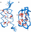
NMR structures of designed proteins GA88 and GB88. (A) NMR ensemble of the 20 final structures for GA88 (residues 6–55) in ribbon representation. (B) NMR ensemble of 20 final structures for GB88 (residues 1–56). The main chain is shown in blue, whereas core and other key side chains are shown in red for both structures.
Fig. 3.
Fig. 3.
Altered local structure in designed proteins. (A) Packing of F52 in GA88 (green) and comparison with A52 in the parent structure, PSD-1 (orange). (B) Backbone and side-chain orientations in the α–β3 loop and β1–β2 loop regions of GB88 (green) compared with the parent GB1 (orange).
Fig. 4.
Fig. 4.
Cα trace backbone superposition of parent and designed protein structures. (A) Structural alignment of GA88 (20-structure ensemble, blue) with PSD-1 (20-structure ensemble, red). (B) Overlay of GB88 (20-structure ensemble, blue) with the x-ray structure of GB1 (red).
Fig. 5.
Fig. 5.
Solvent accessibility in GA88 and GB88. Average values per residue were obtained from 20-structure ensembles for GA88 (open circles) and GB88 (filled circles). Standard deviations are omitted for clarity but were in the following ranges: ±4–20% for highly exposed residues (>50% accessible), ±2–11% for boundary residues (20–50% accessible), and ±0–6% for buried residues (<20% accessible).
Fig. 6.
Fig. 6.
Geometries of nonidentical residues. (A) NMR structure of GA88 highlighting the positions of the seven aa differences (red) between the two sequences. (B) NMR structure of GB88 showing the locations of the seven nonidentities (red). Residues at the disordered N- and C-terminal tails of GA88 have been omitted for clarity. The blue regions represent sequence identity between GA88 and GB88.
Fig. 7.
Fig. 7.
Overlaid 15N-HSQC spectra of GA95 (black) and GB95 (red). Main-chain assignments are indicated and chemical shift index data are included in Table S1 and Table S2. Horizontal lines connect side-chain amide signals. Fifty-three of 56 aa in these two proteins are identical but have different chemical environments reflecting the distinct folds.

Similar articles

Cited by

References

    1. Anfinsen CB. Principles that govern the folding of protein chains. Science. 1973;181:223–230. - PubMed
    1. Lattman EE, Rose GD. Protein folding - what's the question? Proc Natl Acad Sci USA. 1993;90:439–441. - PMC - PubMed
    1. Dahiyat BI, Mayo SL. Probing the role of packing specificity in protein design. Proc Natl Acad Sci USA. 1997;94:10172–10177. - PMC - PubMed
    1. Cordes MH, Walsh NP, McKnight CJ, Sauer RT. Evolution of a protein fold in vitro. Science. 1999;284:325–328. - PubMed
    1. Rose GD, Creamer TP. Protein folding: Predicting predicting. Proteins. 1994;19:1–3. - PubMed

Publication types