. 2008 Sep 23;105(38):14442-6.
doi: 10.1073/pnas.0803678105. Epub 2008 Sep 16.

Protein histidine phosphatase 1 negatively regulates CD4 T cells by inhibiting the K+ channel KCa3.1

Affiliations

Protein histidine phosphatase 1 negatively regulates CD4 T cells by inhibiting the K+ channel KCa3.1

Shekhar Srivastava et al. Proc Natl Acad Sci U S A. .

Abstract

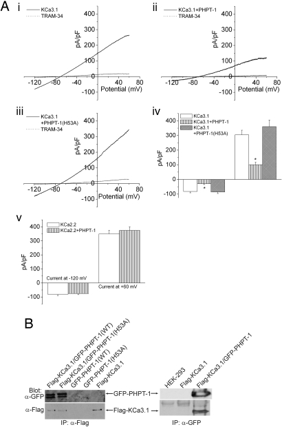
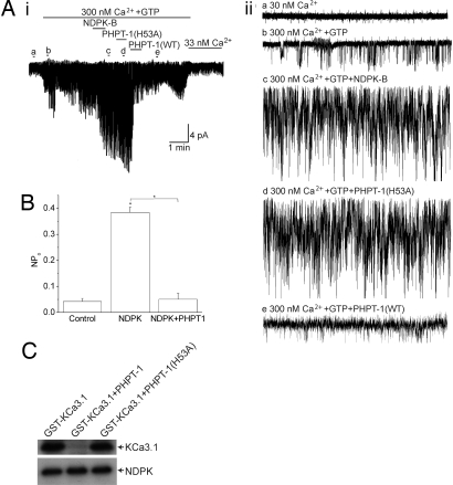
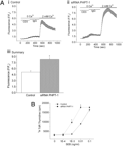
The calcium activated K(+) channel KCa3.1 plays an important role in T lymphocyte Ca(2+) signaling by helping to maintain a negative membrane potential, which provides an electrochemical gradient to drive Ca(2+) influx. We previously showed that nucleoside diphosphate kinase beta (NDPK-B), a mammalian histidine kinase, is required for KCa3.1 channel activation in human CD4 T lymphocytes. We now show that the mammalian protein histidine phosphatase (PHPT-1) directly binds and inhibits KCa3.1 by dephosphorylating histidine 358 on KCa3.1. Overexpression of wild-type, but not a phosphatase dead, PHPT-1 inhibited KCa3.1 channel activity. Decreased expression of PHPT-1 by siRNA in human CD4 T cells resulted in an increase in KCa3.1 channel activity and increased Ca(2+) influx and proliferation after T cell receptor (TCR) activation, indicating that endogenous PHPT-1 functions to negatively regulate CD4 T cells. Our findings provide a previously unrecognized example of a mammalian histidine phosphatase negatively regulating TCR signaling and are one of the few examples of histidine phosphorylation/dephosphorylation influencing a biological process in mammals.


Conflict of interest statement

The authors declare no conflict of interest.

Figures

Fig. 1.
Fig. 1.
Overexpression of PHPT-1 inhibits KCa3.1 channel activity in whole-cell patch-clamp experiments. (A) CHO cells overexpressing KCa3.1 were transfected with GFP, GFP-PHPT-1(WT), or GFP-PHPT-1(H53A), and KCa3.1 channel activity was determined by whole-cell patch-clamp experiments on GFP-positive cells. Shown are current–voltage (i–v) plots of CHO-KCa3.1 cells: control (i), overexpressing GFP-PHPT1(WT) (ii), and overexpressing GFP-PHPT-1(H53A) (iii). Cells in i–iii were inhibited by 1 μM of the selective KCa3.1 blocker TRAM-34 (16). (iv) Bar graph summary of TRAM-34-inhibited currents plotted at −120 and +60 mV (n = 8). (v) Bar graph summary as described in iv showing that overexpression of GFP-PHPT-1(WT) does not inhibit the related calcium-activated potassium channel KCa2.2. (B) PHPT-1 and KCa3.1 coimmunoprecipitate in cells. Flag-KCa3.1 and GFP-PHPT-1(WT) or GFP-PHPT-1(H53A) were transfected into HEK293 cells either alone or together, and cell lysates were then immunoprecipitated (IP) with anti-Flag or anti-GFP antibodies as described (15). The immunoprecipitated proteins were then Western blotted with anti-GFP or anti-Flag antibodies as indicated. Current is represented as pico Amp/pico farad (pA/pF). *, P < 0.05 as compared with control KCa3.1 current. Data displayed as mean ± SEM.
Fig. 2.
Fig. 2.
PHPT-1 directly inhibits KCa3.1 by dephosphorylating H358 in the CT of KCa3.1. (A) I/O patches were isolated from CHO-KCa3.1 cells. Baseline channel activity was first recorded in I/O patches in the absence (i and ii, trace a) or presence of 300 nM Ca2+ and GTP (i and ii, trace b) as described (3). KCa3.1 channels were then activated by the addition of GST-NDPK-B (10 μg/ml) (i and ii, trace c). To determine whether PHPT-1 inhibits KCa3.1 channel activity, His-PHPT-1(H53A) (10 μg/ml) was first added to the same patch (i and ii, trace d), followed by the addition His-PHPT-1(WT) (i and ii, trace e). Aii traces a–e are I/O recordings over 5 sec as indicated. (B) Effect of PHPT-1 on the open channel probability (NPo). Bar graph summary of KCa3.1 NPo from control (trace b), NDPK-B (trace c), and PHPT-1(WT) (trace e); n = 3 patches, P < 0.001. All recordings were at +100 mV. His-PHPT-1(WT), but not His-PHPT-1(H53A), inhibits KCa3.1 channel activity. (C) PHPT-1 dephosphorylates H358 in KCa3.1. To phosphorylate H358 on KCa3.1, Flag-tagged-NDPK-B was immunoprecipitated from transfected HEK293 cell lysates and then incubated with 2.5 μg of GST-KCa3.1(CT) in kinase buffer containing [γ-32P]GTP as described (3). The reaction products were then incubated with 2.5 μg of His-PHPT-1(WT) or His-PHPT-1(H53A) for 30 min at 37°C. Reaction products were then separated by SDS/PAGE and visualized by autoradiography. GST-KCa3.1(CT) and Flag-NDPK-B are indicated. Current represented as pico Amps. *, P < 0.05 as compared with control and as indicated in the figure. Data displayed as mean ± SEM.
Fig. 3.
Fig. 3.
Silencing of PHPT-1 in CD4 T cells by siRNA leads to an increase in KCa3.1 channel activity. Purified CD4 T lymphocytes were transfected with a pool of siRNAs to PHPT-1 (Dharmacon) or a control siRNA by using AMAXA reagents and, after resting overnight, were stimulated with antibodies to CD3 and CD28. Whole-cell patch clamping was performed 48 h after stimulation as described (3). (A) Real-time PCR of PHPT-1 or interleukin 2 from control or CD4+ T cells stimulated with antibodies to CD3 or CD28 for 48 or 72 h (i) or from control or siRNA PHPT-1 transfected cells (ii). The relative amounts of PHPT-1 were standardized against GAPDH. In contrast to mRNA expression of IL-2, T cell stimulation did not lead to an increase in expression of PHPT-1 mRNA. (B) KCa3.1 and Kv1.3 current measured in siRNA control and siRNA PHPT-1 transfected CD4+ T cells. I–V trace of KCa3.1 current from siRNA control (i) and siRNA PHPT-1 transfected (ii) cells. Summary data of TRAM-34 inhibited current at +60 mV from CD4+ T cells transfected with siRNA (–4) PHPT-1 (n = 8–12) (P < 0.001) (iii) or CD4+ T cells transfected with siRNA (–2) or (–4) PHPT-1 (iv). Data are displayed as ± SEM. (v) Kv1.3 current, which was not affected by silencing PHPT-1, was calculated as the remaining current after TRAM-34 treatment. Current is represented as pico Amp/pico farad (pA/pF). *, P < 0.05 as compared with control. Data are displayed as mean ± SEM.
Fig. 4.
Fig. 4.
Silencing of PHPT-1 in CD4+ T cells by siRNA leads to an increase in Ca2+ influx and proliferation. Purified CD4 T cells were transfected with siRNA to PHPT-1 as described in Fig. 3. Cells were then stimulated for 48 h with antibodies to CD3 and CD28 and after resting overnight were loaded with Fluo-4 AM (10 μM). Ca2+ influx was determined by confocal microscopy at 488 nm with images taken every 5 sec after cross-linking with anti-CD3 antibodies (5 mg/ml) as described (17). Average values from 80–100 cells are shown for each series. Ca2+ influx was determined in control (Ai) and siRNA PHPT1 cells (Aii). (Aiii) Bar graph showing fluorescence values from Ai and Aii at peak with 2 mM Ca2+. (B) Purified CD4+ T cells were treated as described in A and, after resting overnight, were plated in 96-well plates with human DC that were activated for 24 h with lipopolysaccharide (100 ng/ml) in a ratio of 10:1 (30,000 CD4+ T cells:3,000 DC) in the presence of increasing concentrations of staphylococcal enterotoxin B (SEB) as described (18). Twenty-four hours after stimulation, cells were pulsed for 8 h with [3H]thymidine, and [3H]thymidine incorporation was assessed by scintillation counting (19). *, P < 0.05 as compared with control. Data are displayed as mean ± SEM.

References

    1. Wulff H, Beeton C, Chandy KG. Potassium channels as therapeutic targets for autoimmune disorders. Curr Opin Drug Discov Devel. 2003;6:640–647. - PubMed
    1. Cahalan MD, Wulff H, Chandy KG. Molecular properties and physiological roles of ion channels in the immune system. J Clin Immunol. 2001;21:235–252. - PubMed
    1. Srivastava S, et al. Histidine phosphorylation of the potassium channel KCa3.1 by nucleoside diphosphate kinase B is required for activation of KCa3.1 and CD4 T cells. Mol Cell. 2006;24:665–675. - PubMed
    1. Ek P, et al. Identification and characterization of a mammalian 14-kDa phosphohistidine phosphatase. Eur J Biochem. 2002;269:5016–5023. - PubMed
    1. Klumpp S, et al. Protein histidine phosphatase: A novel enzyme with potency for neuronal signaling. J Cereb Blood Flow Metab. 2002;22:1420–1424. - PubMed

Publication types

MeSH terms