. 2008 Oct;89(Pt 10):2507-2517.
doi: 10.1099/vir.0.2008/000083-0.

Characterization of hepatitis C RNA-containing particles from human liver by density and size

Affiliations

Characterization of hepatitis C RNA-containing particles from human liver by density and size

Søren U Nielsen et al. J Gen Virol. 2008 Oct.

Abstract

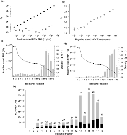
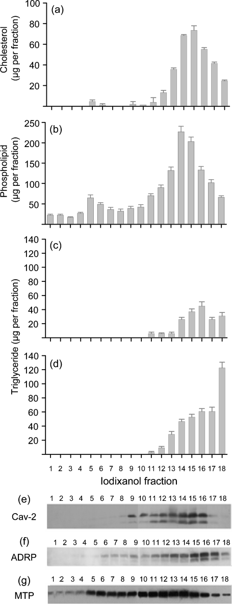
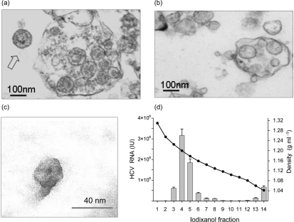
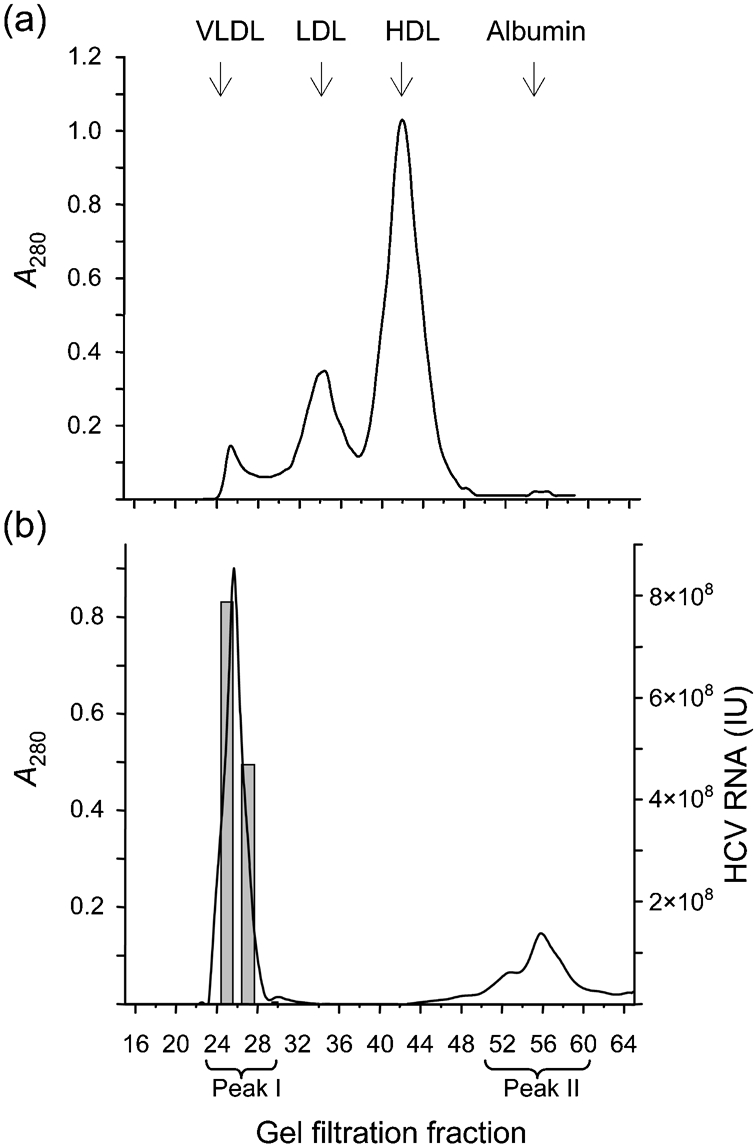
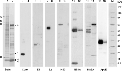
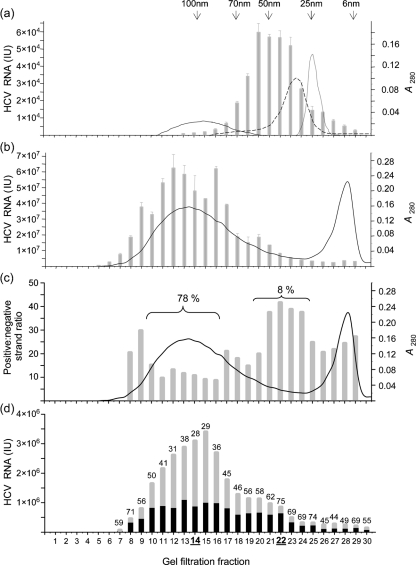
Hepatitis C virus (HCV) particles found in vivo are heterogeneous in density and size, but their detailed characterization has been restricted by the low titre of HCV in human serum. Previously, our group has found that HCV circulates in blood in association with very-low-density lipoprotein (VLDL). Our aim in this study was to characterize HCV RNA-containing membranes and particles in human liver by both density and size and to identify the subcellular compartment(s) where the association with VLDL occurs. HCV was purified by density using iodixanol gradients and by size using gel filtration. Both positive-strand HCV RNA (present in virus particles) and negative-strand HCV RNA (an intermediate in virus replication) were found with densities below 1.08 g ml(-1). Viral structural and non-structural proteins, host proteins ApoB, ApoE and caveolin-2, as well as cholesterol, triglyceride and phospholipids were also detected in these low density fractions. After fractionation by size with Superose gel filtration, HCV RNA and viral proteins co-fractionated with endoplasmic reticulum proteins and VLDL. Fractionation on Toyopearl, which separates particles with diameters up to 200 nm, showed that 78 % of HCV RNA from liver was >100 nm in size, with a positive-/negative-strand ratio of 6 : 1. Also, 8 % of HCV RNA was found in particles with diameters between 40 nm and 70 nm and a positive-/negative-strand ratio of 45 : 1. This HCV was associated with ApoB, ApoE and viral glycoprotein E2, similar to viral particles circulating in serum. Our results indicate that the association between HCV and VLDL occurs in the liver.


Figures

Fig. 1.
Fig. 1.
qRT-PCR assays for positive and negative strand HCV RNA from liver. (a, b) CT values were measured using a dilution series of either synthetic positive strand HCV RNA (a) or negative strand HCV RNA (b) as a template. One primer set and probe was designed to detect positive strand HCV RNA (•) and another primer set was designed with a tagged primer to detect negative strand HCV RNA (○). Each data point represents the mean±the range of values from two determinations. (c, d) The density distribution of positive (c) and negative (d) strand HCV RNA within iodixanol gradients of HCV-infected liver macerate was determined (•). The concentration of RNA is shown by bar graphs which represent the mean±the range of values from two determinations. (e) Immunoprecipitation of HCV in density gradient fractions with antibody to ApoB. The total HCV positive-strand RNA in the pellets and the supernatants (shaded bars) and the HCV RNA associated with ApoB (solid bars) was calculated as a percentage (given above the bars) for each fraction.
Fig. 2.
Fig. 2.
Distribution of lipids (a–d) and proteins (e–g) involved in lipid metabolism within iodixanol density gradient fractions 1–18. Cholesterol (a), phospholipid (b) and triglyceride (d) were measured in each density gradient fraction from liver S6b. Triglyceride was also measured within density gradient fractions from an HCV-negative liver (c). The density distribution of Cav-2 (e), ADRP (f) and MTP (g) in liver S6b was determined by Western blotting.
Fig. 3.
Fig. 3.
Thin-section EM of fraction 15 from iodixanol gradients of HCV-positive liver (a) or a control HCV-negative liver (b). The fraction from the HCV-positive liver had a density of 1.08 g ml−1. The arrow points to an isolated particle which shows internal structure in the form of a pentagon; the diameter of this is 100 nm and the width of the membrane surrounding the particle is 10 nm. (c) Iodixanol fraction 15 was treated with 3 % NP-40 and analysed by negative-staining EM. The density (•) and concentration (bars) of HCV RNA following this treatment (d) indicate that it shifted the density of HCV RNA to 1.2 g ml−1, corresponding to the density of the HCV nucleocapsid (Ishida et al., 2001).
Fig. 4.
Fig. 4.
Separation of HCV RNA-containing particles from liver S6b by gel filtration on Superose 6. (a) The column was calibrated using purified lipoproteins and albumin. Peaks corresponding to VLDL, LDL, HDL and albumin are labelled. (b) Gel-filtration of liver macerate first purified on iodixanol gradients, with density below 1.12 g ml−1. Bars show the titre of positive strand HCV RNA in fractions collected from the column. The line shows the A280 of proteins eluting from the gel filtration column. The analysis was repeated four times with similar results.
Fig. 5.
Fig. 5.
Identification of host and viral proteins after separation by gel filtration. Proteins from gel filtration peaks I and II (Fig. 4b) were analysed by SDS-PAGE (lanes 1 and 2) and Western blotting (lanes 3 to 16). HCV core protein (20 kDa, lane 3) as well as HCV glycoprotein E1 (31 kDa, lane 5) and glycoprotein E2 (62 kDa, lane 7) were detected in peak I but not in peak II (lanes 4, 6 and 8). HCV proteins NS3 (62 kDa, lane 9) and NS4A (7 kDa, lane 11) were also detected in peak I, but not in peak II (lanes 10 and 12). The antibody to HCV NS5A recognized two bands of 55 and 58 kDa in peak I (lane 13). These bands were not present in peak II (lane 14), but cross-reactivity to albumin at 66 kDa was observed. Host lipoprotein ApoE with molecular mass 36 kDa was recognized in peaks I and II (lanes 15 and 16). Proteins A–H in lanes 1 and 2 were excised and identified by MALDI–TOF (Table 2). Lane M, molecular mass marker.
Fig. 6.
Fig. 6.
Analysis of serum and liver macerate from patient S6 by gel filtration on Toyopearl. The gel filtration column was calibrated with latex beads of different sizes (indicated by arrows) as well as with purified chylomicrons, VLDL and LDL. The elution profiles of these proteins are shown as solid, dashed and dotted lines, respectively, in (a). (a) Separation of HCV RNA in serum by gel filtration. Bars show the mean±sd HCV RNA in each fraction. (b) Elution profile of total protein (line). Bars show mean±sd (from three determinations) of HCV RNA from liver macerate with density below 1.12 g ml−1. (c) The ratio of HCV positive strand RNA and HCV negative strand RNA for each fraction. The proportion of HCV RNA within fractions enclosed by brackets is shown as a percentage. (d) Immunoprecipitation of HCV with antibody to ApoB. The total HCV RNA in the pellets and the supernatants (shaded bars) and the HCV RNA associated with ApoB (solid bars) was calculated as a percentage (given above the bars) for each fraction.

Similar articles

Cited by

References

    1. Aizaki, H., Lee, K. J., Sung, V. M. H., Ishiko, H. & Lai, M. M. C. (2004). Characterization of the hepatitis C virus RNA replication complex associated with lipid rafts. Virology 324, 450–461. - PubMed
    1. André, P., Komurian-Pradel, F., Deforges, S., Perret, M., Berland, J. L., Sodoyer, M., Pol, S., Brechot, C., Paranhos-Baccala, G. & Lotteau, V. (2002). Characterization of low- and very-low-density hepatitis C virus RNA-containing particles. J Virol 76, 6919–6928. - PMC - PubMed
    1. Anonymous (2002). Gel filtration. Principles and Methods. Uppsala, Sweden: Amersham Biosciences.
    1. Chang, M., Williams, O., Mittler, J., Quintanilla, A., Carithers, R. L., Perkins, J., Corey, L. & Gretch, D. R. (2003). Dynamics of hepatitis C virus replication in human liver. Am J Pathol 163, 433–444. - PMC - PubMed
    1. Chang, K.-S., Jiang, J., Cai, Z. & Luo, G. (2007). Human apolipoprotein E is required for infectivity and production of hepatitis C virus in cell culture. J Virol 81, 13783–13793. - PMC - PubMed

Publication types

MeSH terms

LinkOut - more resources