Review
. 2008 Nov 4;99(9):1375-9.
doi: 10.1038/sj.bjc.6604662. Epub 2008 Sep 16.

The role of endothelial-to-mesenchymal transition in cancer progression

Affiliations
Review

The role of endothelial-to-mesenchymal transition in cancer progression

S Potenta et al. Br J Cancer. .

Abstract

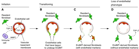
Recent evidence has demonstrated that endothelial-to-mesenchymal transition (EndMT) may have a significant role in a number of diseases. Although EndMT has been previously studied as a critical process in heart development, it is now clear that EndMT can also occur postnatally in various pathologic settings, including cancer and cardiac fibrosis. During EndMT, resident endothelial cells delaminate from an organised cell layer and acquire a mesenchymal phenotype characterised by loss of cell-cell junctions, loss of endothelial markers, gain of mesenchymal markers, and acquisition of invasive and migratory properties. Endothelial-to-mesenchymal transition -derived cells are believed to function as fibroblasts in damaged tissue, and may therefore have an important role in tissue remodelling and fibrosis. In tumours, EndMT is an important source of cancer-associated fibroblasts (CAFs), which are known to facilitate tumour progression in several ways. These new findings suggest that targeting EndMT may be a novel therapeutic strategy, which is broadly applicable not only to cancer but also to various other disease states.


Figures

Figure 1
Figure 1
Stages of EndMT. (A) Endothelial-to-mesenchymal transition may be initiated by autocrine and/or paracrine inflammatory signals originating from within the surrounding tissue, such as TGF-β. Possible sources include resident fibroblasts (green) or immune cells (purple). Alternatively, the endothelium (red) may undergo EndMT in direct response to vascular injury. The vascular basement membrane is likely to be degraded by matrix metalloproteinases (MMPs) derived from local immune cells. (BC) Transitioning endothelial cells (red/green) acquire a migratory phenotype, invade under the vascular basement membrane, and begin to express mesenchymal markers, such as FSP1, while still expressing endothelial markers. (D) Cells that have undergone EndMT (green) have lost their endothelial phenotype. These EndMT-derived cells contribute to the local fibroblast population and are likely to produce various growth factors, such as TGF-β. It is not yet known whether the affected vessels are repopulated, and if they remain functional after resident endothelial cells have departed.
Figure 2
Figure 2
Endothelial-to-mesenchymal transition in cancer and cardiac fibrosis. (A) The Tie2-Cre;ROSA-STOP-lacZ reporter mouse is an important strain for tracking cells of endothelial origin during EndMT. In this mouse, Cre expression is driven by the Tie2 promoter, which is known to be active in endothelial cells. The Cre recombinase acts by permanently excising genomic DNA regions that are flanked by loxP sites (floxed). In this case, Tie2-driven Cre activity removes a floxed stop cassette, thereby allowing lacZ expression to be driven by the constitutive ROSA26R promoter (ROSA) without the need for continued Tie2 activity. (B) During cardiac fibrosis, TGF-β signalling promotes EndMT through Smad3 transcriptional activity. In endothelial cells, TGF-β is known to activate Alk5, which then activates Smad3. However, the role of Alk5 has not been explicitly demonstrated during EndMT in cardiac fibrosis. EndMT was also shown to be inhibited by rhBMP-7 (dashed lines). BMP-7 is known to act through a different set of Smads, namely Smad1, -5, and -8. However, the precise mechanisms whereby BMP-7 inhibits EndMT are not yet known.

References

    1. Arciniegas E, Frid MG, Douglas IS, Stenmark KR (2007) Perspectives on endothelial-to-mesenchymal transition: potential contribution to vascular remodeling in chronic pulmonary hypertension. Am J Physiol Lung Cell Mol Physiol 293: L1–L8 - PubMed
    1. Armstrong EJ, Bischoff J (2004) Heart valve development: endothelial cell signaling and differentiation. Circ Res 95: 459–470 - PMC - PubMed
    1. Armulik A, Abramsson A, Betsholtz C (2005) Endothelial/Pericyte Interactions. Circ Res 97: 512–523 - PubMed
    1. Batlle E, Sancho E, Franci C, Dominguez D, Monfar M, Baulida J, Garcia De Herreros A (2000) The transcription factor snail is a repressor of E-cadherin gene expression in epithelial tumour cells. Nat Cell Biol 2: 84–89 - PubMed
    1. Cano A, Perez-Moreno MA, Rodrigo I, Locascio A, Blanco MJ, del Barrio MG, Portillo F, Nieto MA (2000) The transcription factor snail controls epithelial-mesenchymal transitions by repressing E-cadherin expression. Nat Cell Biol 2: 76–83 - PubMed

Publication types