. 2008;11(4):347-60.
doi: 10.1007/s10456-008-9116-2. Epub 2008 Sep 17.

A role for planar cell polarity signaling in angiogenesis

Affiliations

A role for planar cell polarity signaling in angiogenesis

Pasquale Cirone et al. Angiogenesis. 2008.

Abstract

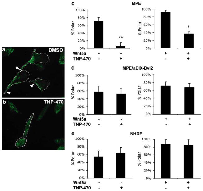
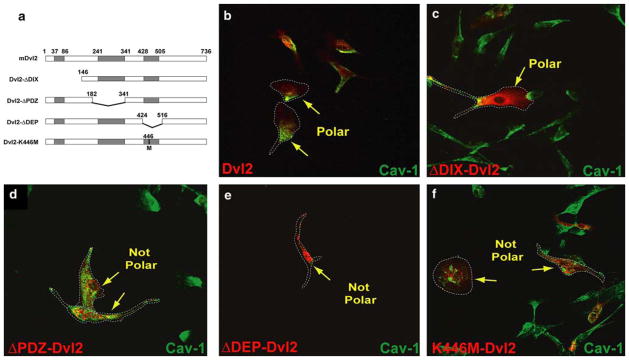
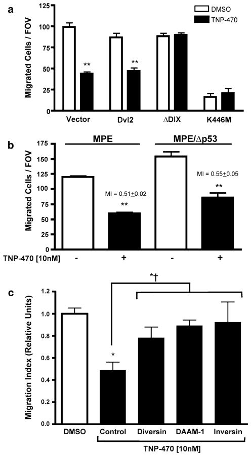
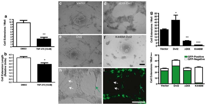
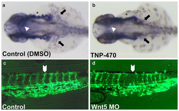
The planar cell polarity (PCP) pathway is a highly conserved signaling cascade that coordinates both epithelial and axonal morphogenic movements during development. Angiogenesis also involves the growth and migration of polarized cells, although the mechanisms underlying their intercellular communication are poorly understood. Here, using cell culture assays, we demonstrate that inhibition of PCP signaling disrupts endothelial cell growth, polarity, and migration, all of which can be rescued through downstream activation of this pathway by expression of either Daam-1, Diversin or Inversin. Silencing of either Dvl2 or Prickle suppressed endothelial cell proliferation. Moreover, loss of p53 rescues endothelial cell growth arrest but not the migration inhibition caused by PCP disruption. In addition, we show that the zebrafish Wnt5 mutant (pipetail (ppt)), which has impaired PCP signaling, displays vascular developmental defects. These findings reveal a potential role for PCP signaling in the coordinated assembly of endothelial cells into vascular structures and have important implications for vascular remodeling in development and disease.


Conflict of interest statement

The authors have declared no competing interests.

Figures

Fig. 1
Fig. 1
TNP-470, a β-catenin-independent Wnt signaling inhibitor, selectively disrupts endothelial cell polarity. a Typical caveolin-1 (green) polar localization to the trailing edge (white arrowheads) of migrating MPE endothelial cells (DMSO control). b Upon the addition of 10 nM TNP-470, caveolin-1 localizes throughout the cytosol (and is no longer polarized to the trailing edge of the cell). c Proportion of MPE cells displaying a polar localization of caveolin-1 (% Polar) in the presence or absence of a Wnt5a-gradient and/or 10 nM TNP-470 (N = 3–5 Dunn chemotaxis chambers, each N = 12 fields of view (FOV)). d Depolarizing activity of TNP-470 is rescued upon activation of β-catenin-independent signaling by ΔDIX-Dvl2 (N = 3–5 Dunn chemotaxis chambers, each N = 12 FOV) (P >0.05). e Polarization of caveolin-1 in NHDF cells in Dunn chemotaxis chambers. The addition of TNP-470 did not disrupt caveolin-1 localization in NHDF cells (P >0.05). Dashed lines encircle cells to facilitate their visualization
Fig. 2
Fig. 2
Dvl2 mutants disrupt endothelial cell polarity. a Diagram of HA-Dvl2 constructs in pCMV5 (HA-tags on the N-terminal of each construct are not depicted). Each of the functional domains have been individually deleted. In addition, one construct was generated through the introduction of a point mutation to yield the PCP-inhibitory K446M-Dvl2 construct. Caveolin-1 localization (b–f, green) in MPE cells expressing various HA-Dvl2 (red) constructs: b Dvl2, c ΔDIX-Dvl2, d ΔPDZ-Dvl2, e ΔDEP-Dvl2 and f K446M-Dvl2. Normal asymmetrical caveolin-1 localization was observed in Dvl2 and ΔDIX-Dvl2-expressing cells but was either diminished or mis-localized in cells expressing the others constructs. The minimal K446M point-mutation in Dvl2 was sufficient to bring about this phenotype. Dashed lines encircle cells to facilitate their visualization
Fig. 3
Fig. 3
A PCP-deficient point-mutation form of Dvl2 (K446M-Dvl2) disrupts endothelial cell proliferation. a Western blot of Dvl2 constructs in MPE whole cell lysates. b Expression of WT or DIX-deleted forms of Dvl2 did not affect MPE cell proliferation. MPE cells that expressed either ΔPDZ or ΔDEP mutants were not viable and were therefore not included in this assay. However, endothelial cells expressing K446M-Dvl2 were predominantly cytostatic (***; P <0.001, N = 4/group, triplicate experiments)
Fig. 4
Fig. 4
PCP signaling inhibition disrupts endothelial cell migration in a p53-indpendant manner. a Migration of MPE cells transiently co-transfected with various Dvl2/GFP constructs (**, P <0.01). b TNP-470 disrupts endothelial cell migration independently of p53 status. The difference between migration indices (MI) was not statistically significant (P >0.05). c Expression of various PCP mediators, including Diversin, DAAM-1 and Inversin rescue from TNP-470-Induced Non-canonical signaling inhibition. TNP-470 treatment significantly inhibited migration of MPE cells (*; P <0.05). Migration indices were determined for different positive PCP mediators for protection against TNP-470 and were statistically not distinguishable from the DMSO control (P >0.05) but meaningfully greater than TNP-470 treatment (*<; P <0.05)
Fig. 5
Fig. 5
Both over-activation and inhibition of PCP signaling disrupt endothelial cell network formation. (a–b) TNP-470 inhibits the initiation of endothelial network formation in vitro. MPE cells were plated onto Matrigel with or without TNP-470. a The number of extensions formed was drastically reduced in the presence of 10-nM TNP-470. b Cell-extension length was reduced by approximately 25% with 10-nM TNP-470. c–f Networks formed by MPE cells expressing various Dvl2 constructs (as labeled). g Quantification of cellular extensions formed depicted in (c–f) (N = 8/group). h–j MPE cells co-expressing Dvl2 mutations and GFP were mixed with non-transfected MPE (mosaic analysis) and incubated on Matrigel. h Phase contrast and i green fluorescence images of MPE cells within the mosaic analysis. Cell extensions were thereafter scored as GFP-positive (green arrows) or GFP-negative (white arrows) if cell extensions were devoid of GFP-positive cells. j Quantification of extensions formed in the mosaic analysis (N = 8/group). Values within each of the groups (GFP+ vs. GFP) were statistically different (P <0.01). The non-mutant Dvl2 group displayed more extensive networks over other test conditions (P <0.05). The quantities were significantly different for the Dvl2-autonomous (increase, P <0.05) and K446M-Dvl2-autonomous (decrease, P <0.01) groups. All scale bars 200 μm
Fig. 6
Fig. 6
Wnt5-deficient (ppt) zebrafish display aberrant angiogenesis in vivo. a–b Tg(fli:EGFP) expression (28–30 hpf) as detected by whole mount. An in situ Fli probe shows ordered intersegmental vessel (IS) development in wt-like embryos (a, arrow) as well as proper development of the dorsal aorta (DA), and posterior cardinal vein (PCV) (arrowhead). b Similarly staged ppt mutants display disrupted angiogenesis with defective IS vessel formation (arrow) and reduced DA/PCV expression (arrowhead). c–d Fli-GFP expression in vivo. WT (c) and ppt (d) expression of Fli-GFP. Loss of Wnt5 results in disruption of the Duct of Cuvier/CCV. (e–f) Live in vivo Fli-GFP images of 48 hpf embryos. Irregular vessel formation is seen in ppt relative to WT control. g–h Whole mount staining of WT and ppt embryo head regions. g Robust vasculature can be observed in the anterior and middle cerebral veins within WT embryos. These vessels are diminished and aberrantly patterned within ppt embryos (h)
Fig. 7
Fig. 7
Vascular defects due to chemical inhibition of PCP signaling mimic those of ppt zebrafish. a–b Tg(fli:EGFP) expression (28–30 hpf) as detected by whole mount. Relative to WT (a) vasculature, TNP-470-treated animals (b) displayed disrupted vessel growth (asterisks), disrupted common cardinal vein formation (arrows) and markedly diminished middle cerebral vein (arrowheads). c–d Wnt5 MO-injected embryos evaluated for vasculature defects. Fluorescent imaging of Control (c) and Wnt MO-injected embryos (d), lateral view of the tail. Wnt5-MO injection (d) disrupts intersegmental angiogenesis in Tg(fli:EGFP)

References

    1. Liebner S, Cavallaro U, Dejana E. The multiple languages of endothelial cell-to-cell communication. Arterioscler Thromb Vasc Biol. 2006;26:1431–1438. doi: 10.1161/01.ATV.0000218510. 04541.5e. - DOI - PubMed
    1. Fanto M, McNeill H. Planar polarity from flies to vertebrates. J Cell Sci. 2004;117:527–533. doi: 10.1242/jcs.00973. - DOI - PubMed
    1. Gong Y, Mo C, Fraser SE. Planar cell polarity signalling controls cell division orientation during zebrafish gastrulation. Nature. 2004;430:689–693. doi: 10.1038/nature02796. - DOI - PubMed
    1. Heisenberg CP, Tada M, Rauch GJ, Saude L, Concha ML, Geisler R, et al. Silberblick/Wnt11 mediates convergent extension movements during zebrafish gastrulation. Nature. 2000;405:76–81. doi: 10.1038/35011068. - DOI - PubMed
    1. Myers DC, Sepich DS, Solnica-Krezel L. Convergence and extension in vertebrate gastrulae: cell movements according to or in search of identity? Trends Genet. 2002;18:447–455. doi: 10.1016/S0168-9525(02)02725-7. - DOI - PubMed

Publication types

MeSH terms