

Regulation of brain-derived neurotrophic factor-mediated transcription of the immediate early gene Arc by intracellular calcium and calmodulin
- PMID: 18798281
- PMCID: PMC2628963
- DOI: 10.1002/jnr.21863
Regulation of brain-derived neurotrophic factor-mediated transcription of the immediate early gene Arc by intracellular calcium and calmodulin
Abstract
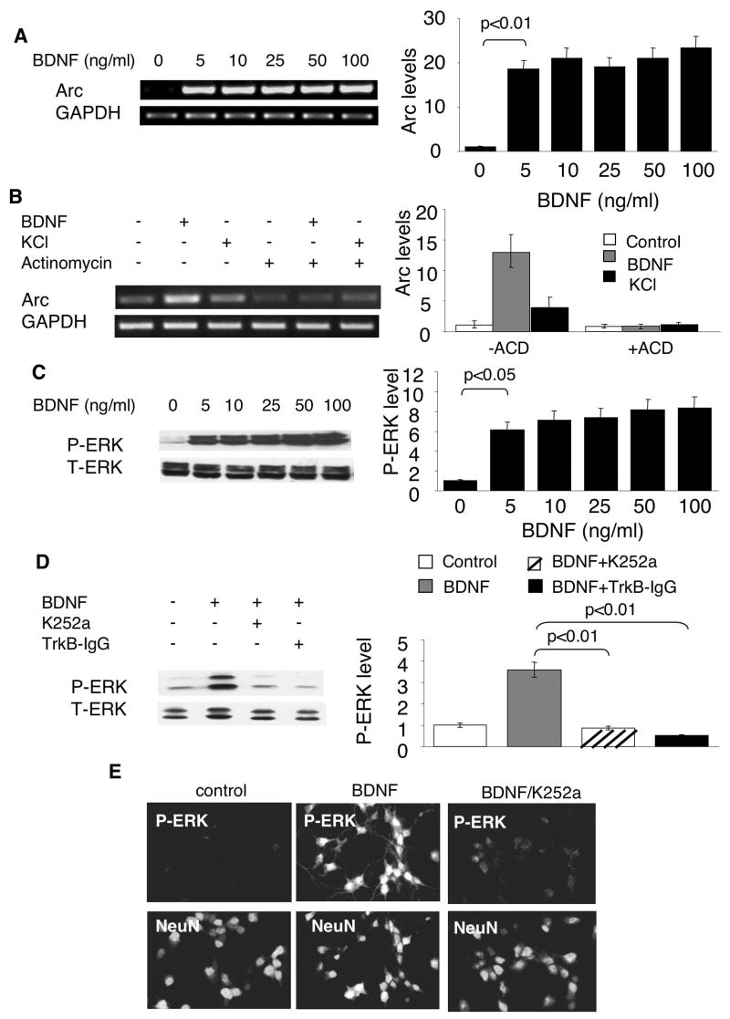
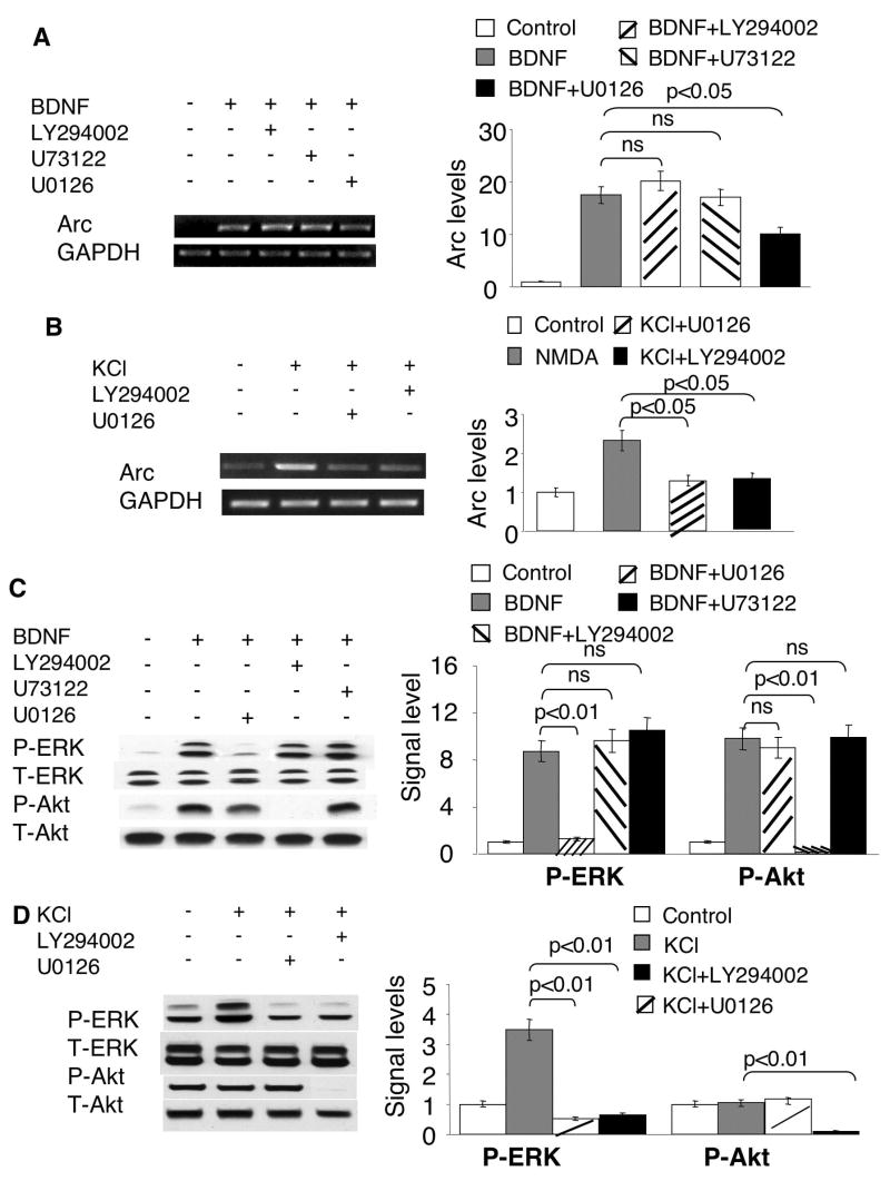
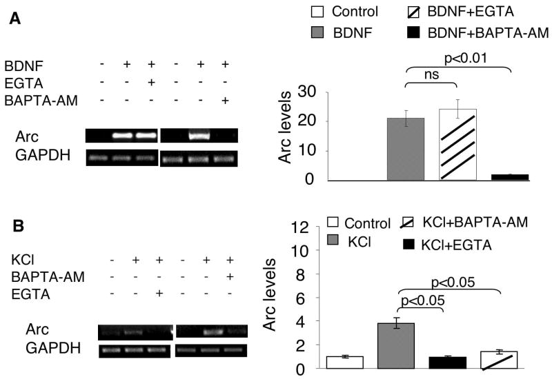
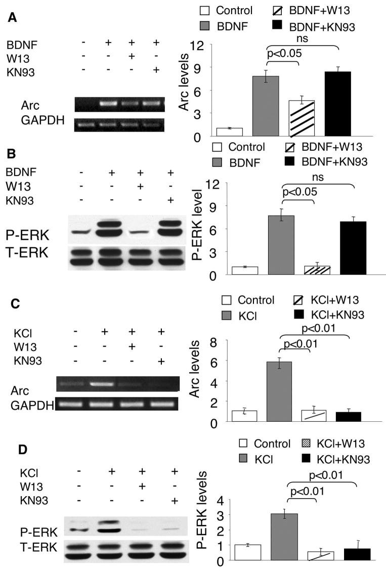
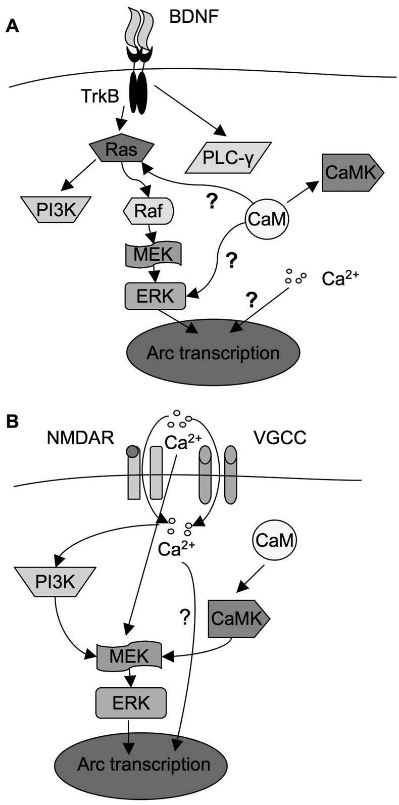
The induction of the immediate early gene Arc is strongly implicated in synaptic plasticity. Although the role of ERK has been demonstrated, the regulation of Arc expression is largely unknown. In this study, we investigated the major signaling pathways underlying brain-derived neurotrophic factor (BDNF)-mediated Arc transcription in cultured cortical neurons. The BDNF-stimulated Arc transcription was regulated solely by the Ras-Raf-MAPK signaling through ERK, but not by phosphoinositide 3-kinase (PI3K) and PLC-gamma activities. Although it was demonstrated that BDNF might promote calcium entry through calcium channels and NMDA receptors, chelating extracellular calcium with EGTA failed to block Arc transcription. In contrast, chelating intracellular calcium ([Ca(2+)](i)) by BAPTA-AM abolished BDNF-mediated Arc up-regulation. Surprisingly, BAPTA-AM did not block ERK activation, indicating that [Ca(2+)](i) and Ras-Raf-MAPK are not coupled, and the activation of ERK alone is not sufficient to up-regulate Arc transcription. Moreover, we found that inhibition of calmodulin (CaM) by W13 blocked both Arc transcription and ERK activation, revealing a Ca(2+)-independent function of CaM. These data suggested novel functions of [Ca(2+)](i) and CaM in BDNF signaling. Comparison of the Arc transcription profiles between Ca(2+)-stimulated and BDNF-stimulated neurons demonstrated that the regulatory mechanisms were distinctively tailored to the complex features of neuronal activity. Specifically, PI3K and CaM-dependent protein kinase (CaMK) activity were required for Ca(2+)-stimulated Arc transcription through regulating ERK signaling. Such cross-talks between PI3K, CaMK, and ERK was absent in BDNF-stimulated neurons.
2008 Wiley-Liss, Inc.
Figures


Similar articles
-
Intracellular calcium and calmodulin link brain-derived neurotrophic factor to p70S6 kinase phosphorylation and dendritic protein synthesis.J Neurosci Res. 2010 May 15;88(7):1420-32. doi: 10.1002/jnr.22321. J Neurosci Res. 2010. PMID: 20029971 Free PMC article.
-
NMDA-mediated and self-induced bdnf exon IV transcriptions are differentially regulated in cultured cortical neurons.Neurochem Int. 2009 May-Jun;54(5-6):385-92. doi: 10.1016/j.neuint.2009.01.006. Neurochem Int. 2009. PMID: 19418634 Free PMC article.
-
The basal level of intracellular calcium gates the activation of phosphoinositide 3-kinase-Akt signaling by brain-derived neurotrophic factor in cortical neurons.J Neurochem. 2008 Aug;106(3):1259-74. doi: 10.1111/j.1471-4159.2008.05478.x. Epub 2008 May 12. J Neurochem. 2008. PMID: 18485103 Free PMC article.
-
BDNF-induced local protein synthesis and synaptic plasticity.Neuropharmacology. 2014 Jan;76 Pt C:639-56. doi: 10.1016/j.neuropharm.2013.04.005. Epub 2013 Apr 16. Neuropharmacology. 2014. PMID: 23602987 Review.
-
BDNF mechanisms in late LTP formation: A synthesis and breakdown.Neuropharmacology. 2014 Jan;76 Pt C:664-76. doi: 10.1016/j.neuropharm.2013.06.024. Epub 2013 Jul 2. Neuropharmacology. 2014. PMID: 23831365 Review.
Cited by
-
Short-term striatal gene expression responses to brain-derived neurotrophic factor are dependent on MEK and ERK activation.PLoS One. 2009;4(4):e5292. doi: 10.1371/journal.pone.0005292. Epub 2009 Apr 23. PLoS One. 2009. PMID: 19390590 Free PMC article.
-
Role of Brain Derived Neurotrophic Factor and Related Therapeutic Strategies in Central Post-Stroke Pain.Neurochem Res. 2024 Sep;49(9):2303-2318. doi: 10.1007/s11064-024-04175-z. Epub 2024 Jun 10. Neurochem Res. 2024. PMID: 38856889 Review.
-
Novelty exposure overcomes foot shock-induced spatial-memory impairment by processes of synaptic-tagging in rats.Proc Natl Acad Sci U S A. 2012 Jan 17;109(3):953-8. doi: 10.1073/pnas.1114198109. Epub 2012 Jan 3. Proc Natl Acad Sci U S A. 2012. PMID: 22215603 Free PMC article.
-
Gene expression changes in the medial prefrontal cortex and nucleus accumbens following abstinence from cocaine self-administration.BMC Neurosci. 2010 Feb 26;11:29. doi: 10.1186/1471-2202-11-29. BMC Neurosci. 2010. PMID: 20187946 Free PMC article.
-
Isoxazole Alters Metabolites and Gene Expression, Decreasing Proliferation and Promoting a Neuroendocrine Phenotype in β-Cells.ACS Chem Biol. 2016 Apr 15;11(4):1128-36. doi: 10.1021/acschembio.5b00993. Epub 2016 Feb 10. ACS Chem Biol. 2016. PMID: 26828310 Free PMC article.
References
-
- Bramham CR. Control of synaptic consolidation in the dentate gyrus: mechanisms, functions, and therapeutic implications. Prog Brain Res. 2007;163:453–471. - PubMed
-
- Chen X, Garelick MG, Wang H, Lil V, Athos J, Storm DR. PI3 kinase signaling is required for retrieval and extinction of contextual memory. Nat Neurosci. 2005;8(7):925–931. - PubMed
-
- Della Rocca GJ, Mukhin YV, Garnovskaya MN, Daaka Y, Clark GJ, Luttrell LM, Lefkowitz RJ, Raymond JR. Serotonin 5-HT1A receptor-mediated Erk activation requires calcium/calmodulin-dependent receptor endocytosis. J Biol Chem. 1999;274(8):4749–4753. - PubMed
Publication types
MeSH terms
Substances
Grants and funding
LinkOut - more resources
Full Text Sources
Research Materials
Miscellaneous
