

Bone marrow-derived immune cells mediate sensitization to liver injury in a myeloid differentiation factor 88-dependent fashion
- PMID: 18798338
- PMCID: PMC7043384
- DOI: 10.1002/hep.22557
Bone marrow-derived immune cells mediate sensitization to liver injury in a myeloid differentiation factor 88-dependent fashion
Abstract
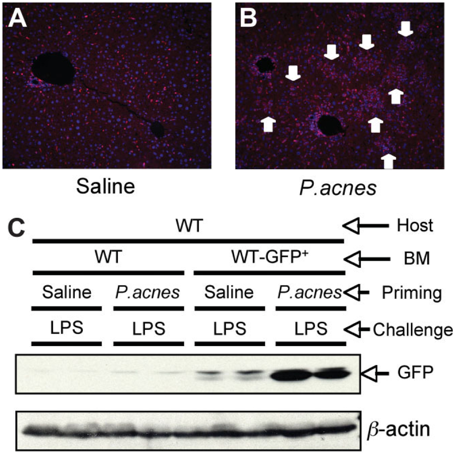
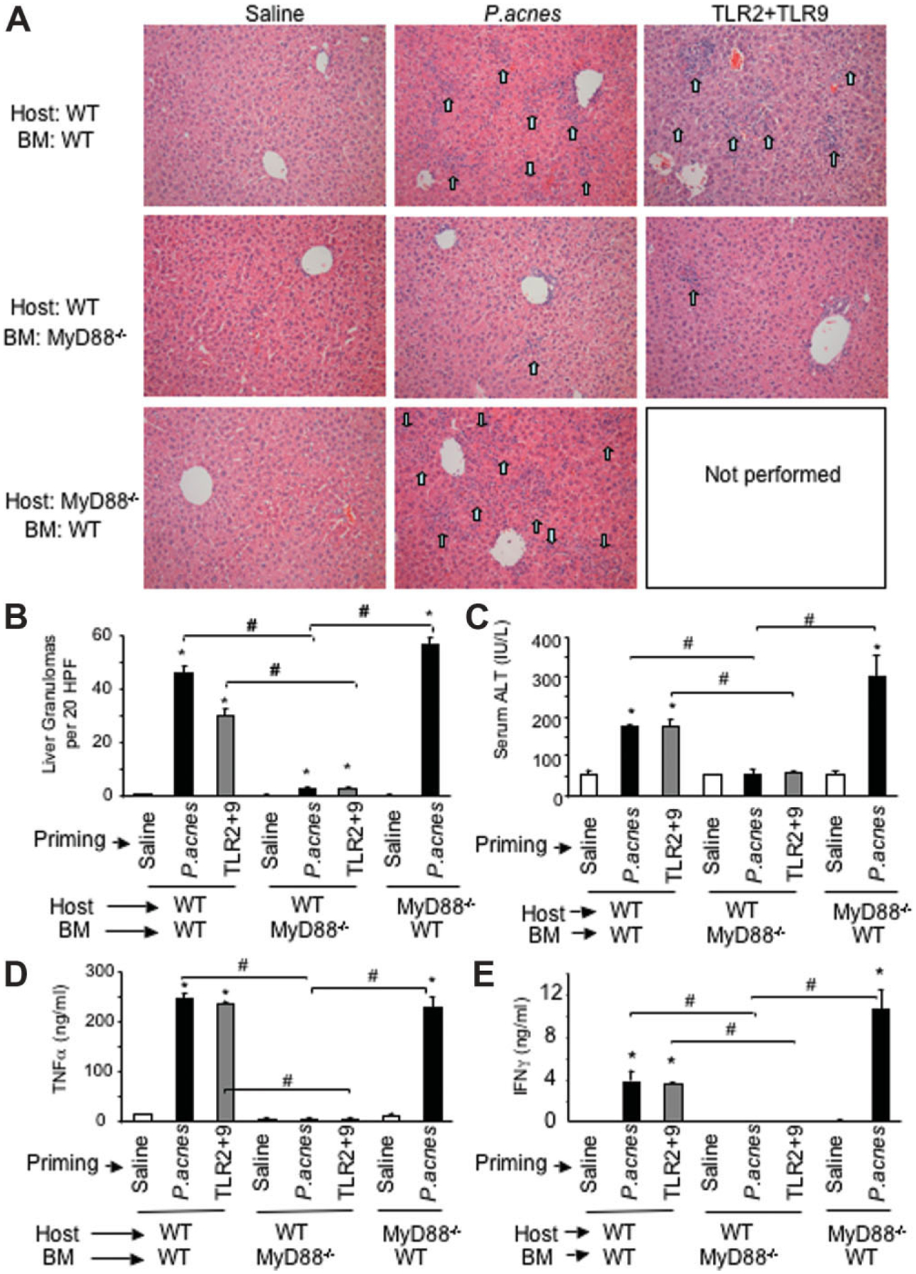
Toll-like receptors (TLRs) expressed on both immune cells and hepatocytes recognize microbial danger signals and regulate immune responses. Previous studies showed that TLR9 and TLR2 mediate Propionibacterium acnes-induced sensitization to lipopolysaccharide-triggered acute liver injury in mice. Ligand-specific activation of TLR2 and TLR9 are dependent on the common TLR adaptor, myeloid differentiation factor 88 (MyD88). Here, we dissected the role of MyD88 in parenchymal and bone marrow (BM)-derived cells in liver sensitization. Using chimeric mice with green fluorescent protein-expressing BM cells, we identified that P. acnes-induced liver inflammatory foci are of BM origin. Chimeras with MyD88-deficient BM showed no inflammatory foci after P. acnes or TLR2+TLR9 challenge, suggesting that recruitment of inflammatory cells to the liver required MyD88 expression in BM-derived cells. Further, selective MyD88 deficiency in parenchymal cells in mice with wild-type BM failed to prevent inflammatory cell infiltration. These results demonstrate that MyD88 in immune cells rather than in liver parenchymal cells plays an important role in inflammatory cell recruitment and liver injury.
Conflict of interest statement
Potential conflict of interest: Nothing to report.
Figures
References
-
- Williams R. Classification, etiology, and considerations of outcome in liver failure. Semin Liver Dis 1996;16:343–348. - PubMed
-
- Pasare C, Medzhitov R. Toll-like receptors: linking innate and adaptive immunity. Adv Exp Med Biol 2005;560:11–18. - PubMed
-
- Zarember KA, Godowski PJ. Tissue expression of human Toll-like receptors and differential regulation of Toll-like receptor mRNAs in leukocytes in response to microbes, their products, and cytokines. J Immunol 2002; 168:554–561. - PubMed
-
- Szabo G, Dolganiuc A, Mandrekar P. Pattern recognition receptors: a contemporary view on liver diseases. HEPATOLOGY 2006;44:287–298. - PubMed
-
- Kawai T, Akira S. TLR signaling. Semin Immunol 2007;19:24–32. - PubMed
MeSH terms
Substances
Grants and funding
LinkOut - more resources
Full Text Sources
Medical