. 2009 Jan;21(1):85-93.
doi: 10.1111/j.1365-2982.2008.01185.x. Epub 2008 Sep 16.

Apoptotic cell death of human interstitial cells of Cajal

Affiliations

Apoptotic cell death of human interstitial cells of Cajal

S J Gibbons et al. Neurogastroenterol Motil. 2009 Jan.

Abstract

Interstitial cells of Cajal (ICC) are specialized mesenchyme-derived cells that regulate contractility and excitability of many smooth muscles with loss of ICC seen in a variety of gut motility disorders. Maintenance of ICC numbers is tightly regulated, with several factors known to regulate proliferation. In contrast, the fate of ICC is not established. The aim of this study was to investigate whether apoptosis plays a role in the regulation of ICC numbers in the normal colon. ICC were identified by immunolabelling for the c-Kit receptor tyrosine kinase and by electron microscopy. Apoptosis was detected in colon tissue by immunolabelling for activated caspase-3, terminal dUTP nucleotide end labelling and by ultrastructural changes in the cells. Apoptotic ICC were identified and counted in double-labelled tissue sections. They were identified in all layers of the colonic muscle. In the muscularis propria 1.5 +/- 0.2% of ICC were positive for activated caspase-3 and in the circular muscle layer 2.1 +/- 0.9% of ICC were positive for TUNEL. Apoptotic ICC were identified by electron microscopy. Apoptotic cell death is a continuing process in ICC. The level of apoptosis in ICC in healthy colon indicates that these cells must be continually regenerated to maintain intact networks.


Figures

Figure 1
Figure 1. Activated caspase-3 positive interstitial cells of Cajal (ICC) in the human colon
Representative activated caspase-3 (red staining and red fluorescence indicated by arrowheads) and c-Kit positive ICC (brown staining) are shown from sections of human colon with counterstained nuclei (blue). The left panels in a and b show white light illuminated, bright field images, the middle panels show the fluorescent structures in the same field (Ex 560 nm, Em 645), the right panels are merged images. a) example of an activated caspase-3 positive ICC in circular muscle from a 40 year old male; b) another example of an activated caspase-3 positive ICC in the myenteric plexus region of the colon of a 45 year old female. Arrows in b and c indicate activated caspase-3 positive cells that are not ICC. The asterisks indicate two ICC that are negative for activated caspase-3. Scale bars = 25 µm. c) Numbers of activated caspase-3 positive ICC expressed as the number of apoptotic ICC per mm3 of total muscle volume. The values obtained from the individual patient samples are labeled with the ages of the patients. The mean ± SEM for the 7 patients is shown on the right.
Figure 2
Figure 2. TUNEL positive interstitial cells of Cajal (ICC) in the human colon
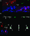
Representative images of TUNEL positive and TUNEL negative ICC from sections of human colon. a) A TUNEL-positive ICC (arrowhead) immediately adjacent to a TUNEL-negative ICC in the myenteric plexus region (arrow) and close to other TUNEL-negative ICC (arrow). The top left image in each panel shows Kit immunostaining (red), the top right TUNEL (green), the bottom left DAPI (blue) and the bottom right the merged images. b) 3D volume rendered images of the same image as (a) rotated 90 degrees to show localization of the TUNEL-positive nucleus (green) within the c-Kit immunoreactive ICC (transparent white). c) a TUNEL positive ICC from the circular muscle. The top left image in each panel shows Kit immunostaining (red), the top right TUNEL (green), the bottom left DAPI (blue) and the bottom right the merged images. d) 3D volume rendered image of the same image as (c) showing localization of the TUNEL image (green) within the c-Kit positive structures (transparent white) shown in two planes 90 degrees rotated from each other. Examples of TUNEL-positive cells are from a 45 year old female. e) Numbers of TUNEL-positive ICC expressed as the % of total number of ICC in the circular muscle layer of each section. On the left is shown the values obtained from individual patient samples labeled with the ages of the patients; on the right is the mean ± SEM for the 9 patients. f) Numbers of all TUNEL positive cells and TUNEL positive ICC in the circular muscle layer. The numbers of TUNEL-positive cells per field obtained from individual tissue samples are shown labeled with the ages of the patients together with the mean ± SEM for all the tissues in a separate column.
Figure 3
Figure 3
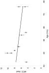
Number of circular muscle interstitial cells of Cajal (ICC)/field in the normal human colon plotted against age of the patients. Data are the mean ± SEM obtained by counting ICC in an average of 132 fields per patient. Each field was 0.95 mm2 and 6 to 10 tissue sections were counted for each tissue specimen. The fit to the data did not show a significant change in the number of ICC with age (r2 = 0.14, P>0.05).
Figure 4
Figure 4
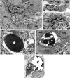
Electron micrographs of interstitial cells of Cajal (ICC) from normal human colon. a) Two ICC in the circular muscle layer in contact with smooth muscle cells. These ICC are also close to a bundle of nerve fibers. One of these ICC has the classical features of ICC while the second one (lower ICC) also has several cisternae of rough endoplasmic reticulum. b) An ICC also showing fibroblast-like features with an extended rough endoplasmic reticulum and a large Golgi apparatus. This cell is in contact with a smooth muscle cell (arrow). c) A detail of a peg-and-socket junction (asterisk) between an ICC and a smooth muscle cell. d and e: apoptotic ICC. In d, the nucleus of the ICC (asterisk) is filled with a condensed chromatin and the cytoplasm is empty. Arrows indicate contact areas between the ICC and neighboring smooth muscle cells. In e, an ICC (asterisks) whose nucleus shows intensely heterochromatic blocks (white asterisk) immersed in a clear matrix. The nuclear envelope is discontinuous and the cytoplasm contains only some lysosomes and few cisternae of the endoplasmic reticulum. The arrow indicates a contact area between the ICC and one smooth muscle cell. In f, an apoptotic smooth muscle cell. Scale bar: a, 1.6 µm; b, 2 µm; c, 0.8 µm; d, 1 µm; e, 1.3 µm; f, 1.3 µm. Interstitial cell of Cajal: ICC, smooth muscle cell, SMC, nerve fiber NF.

References

    1. Thomsen L, Robinson TL, Lee JC, et al. Interstitial cells of Cajal generate a rhythmic pacemaker current. Nature Med. 1998;4:848–51. - PubMed
    1. Ward SM, Burns AJ, Torihashi S, Sanders KM. Mutation of the proto-oncogene c-kit blocks development of interstitial cells and electrical rhythmicity in murine intestine. J Physiol. 1994;480:91–7. - PMC - PubMed
    1. Huizinga JD, Thuneberg L, Kluppel M, Malysz J, Mikkelsen HB, Bernstein A. W/kit gene required for interstitial cells of Cajal and for intestinal pacemaker activity. Nature. 1995;373:347–9. - PubMed
    1. Burns AJ, Lomax AE, Torihashi S, Sanders KM, Ward SM. Interstitial cells of Cajal mediate inhibitory neurotransmission in the stomach. Proc Nat Acad Sci USA. 1996;93:12008–13. - PMC - PubMed
    1. Ward SM, Beckett EA, Wang X, Baker F, Khoyi M, Sanders KM. Interstitial cells of Cajal mediate cholinergic neurotransmission from enteric motor neurons. J Neuroscience. 2000;20:1393–403. - PMC - PubMed

Publication types