. 2008 Nov;82(22):11398-409.
doi: 10.1128/JVI.02654-07. Epub 2008 Sep 17.

Residues in the stalk domain of the hendra virus g glycoprotein modulate conformational changes associated with receptor binding

Affiliations

Residues in the stalk domain of the hendra virus g glycoprotein modulate conformational changes associated with receptor binding

Kimberly A Bishop et al. J Virol. 2008 Nov.

Abstract

Hendra virus (HeV) is a member of the broadly tropic and highly pathogenic paramyxovirus genus Henipavirus. HeV is enveloped and infects cells by using membrane-anchored attachment (G) and fusion (F) glycoproteins. G possesses an N-terminal cytoplasmic tail, an external membrane-proximal stalk domain, and a C-terminal globular head that binds the recently identified receptors ephrinB2 and ephrinB3. Receptor binding is presumed to induce conformational changes in G that subsequently trigger F-mediated fusion. The stalk domains of other attachment glycoproteins appear important for oligomerization and F interaction and specificity. However, this region of G has not been functionally characterized. Here we performed a mutagenesis analysis of the HeV G stalk, targeting a series of isoleucine residues within a hydrophobic alpha-helical domain that is well conserved across several attachment glycoproteins. Nine of 12 individual HeV G alanine substitution mutants possessed a complete defect in fusion-promotion activity yet were cell surface expressed and recognized by a panel of conformation-dependent monoclonal antibodies (MAbs) and maintained their oligomeric structure. Interestingly, these G mutations also resulted in the appearance of an additional electrophoretic species corresponding to a slightly altered glycosylated form. Analysis revealed that these G mutants appeared to adopt a receptor-bound conformation in the absence of receptor, as measured with a panel of MAbs that preferentially recognize G in a receptor-bound state. Further, this phenotype also correlated with an inability to associate with F and in triggering fusion even after receptor engagement. Together, these data suggest the stalk domain of G plays an important role in the conformational stability and receptor binding-triggered changes leading to productive fusion, such as the dissociation of G and F.


Figures

FIG. 1.
FIG. 1.
Sequence alignment of the stalk regions in selected paramyxovirus attachment proteins. (A) Partial sequence alignment of the HeV G stalk domain with that of other paramyxovirus attachment proteins. Asterisks indicate the motif of conserved isoleucine, leucine, and valine residues. (B) That particular motif in the stalk of HeV G consisting solely of isoleucines. Large, boldfaced residues are those which were mutated to alanine and subsequently assessed for fusion promotion activity in the current study. The I118 residue that we were unable to mutate is shown in plain, unbolded text and underlined.
FIG. 2.
FIG. 2.
Fusion promotion activity of HeV G glycoprotein stalk mutants. The various HeV G alanine mutants or WT HeV G were coexpressed with HeV F and assayed for their ability to promote cell-cell fusion when mixed with receptor-positive 293T or PCI-13 cells in a quantitative, vaccinia virus-based fusion assay as described in Materials and Methods. HeLa-USU cells, which are receptor-negative for henipaviruses, served as a negative control. The means of two independent experiments are shown. Error bars represent the ranges. This experiment has been conducted more than five times, and a representative result is shown.
FIG. 3.
FIG. 3.
Cell surface expression of HeV G glycoprotein stalk mutants. Proteins on the surfaces of HeLa-USU cells transiently expressing HeV G alanine mutants or WT HeV G were biotin labeled at 4°C. Lysates were prepared, and biotin-labeled proteins were immunoprecipitated with avidin-agarose beads, subjected to SDS-PAGE, and immunoblotted with G-specific antisera as described in Materials and Methods. Total cell lysates were also probed with polyclonal G-specific antisera for comparison (control). Arrows point to the two species of HeV G.
FIG. 4.
FIG. 4.
Receptor binding by HeV G glycoprotein stalk mutants. The various alanine mutants or WT HeV G were transiently expressed in HeLa-USU cells and subjected to coprecipitation with s-tagged human ephrinB2 followed by S-agarose beads or Fc-tagged human ephrinB3 followed by protein G beads. Each lysate was also directly precipitated with polyclonal G-specific antiserum followed by protein G beads for comparison (control). Precipitated proteins were analyzed by SDS-PAGE followed by Western blotting with polyclonal G-specific antiserum.
FIG. 5.
FIG. 5.
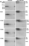
Oligomerization of HeV G glycoprotein stalk mutants. Various alanine mutants or WT HeV G were transiently expressed in HeLa-USU cells, metabolically labeled, and chased as described in Materials and Methods. Each lysate was layered onto a continuous 5 to 20% sucrose gradient and centrifuged at 40,000 rpm for 20 h at 4°C. Then, each gradient was fractionated and immunoprecipitated with polyclonal G-specific antiserum and analyzed by SDS-PAGE under reducing and nonreducing conditions followed by autoradiography. The last two fractions of the I112A mutant are not shown for the sake of organizational clarity. The different species of G, monomer (m), dimer (d), and tetramer, are indicated on the right.
FIG. 6.
FIG. 6.
Complex oligosaccharide addition to HeV G glycoprotein stalk mutants. The apparent higher-molecular-weight species of fusion-defective G stalk mutants was explored. (A) G alanine mutants or WT HeV G were expressed in HeLa-USU cells and lysates were prepared as described in Materials and Methods. G-containing lysates were immunoprecipitated with polyclonal G-specific antiserum followed by protein G beads. Precipitated proteins were treated with PNGase F or Endo HF at 37°C for 0, 10, or 60 min, and the reactions were analyzed by SDS-PAGE and Western blotting with polyclonal G-specific antiserum. (B) Schematic of the HeV G glycoprotein, illustrating the location of eight potential N-linked glycosylation sites (asterisks) in reference to the stalk domain of G. (C) Effect of individual glycosylation site deletions on fusion promotion ability of G, assessed in a quantitative, vaccinia virus-based cell fusion assay as described in Materials and Methods. The reactions were conducted in duplicate wells using receptor-positive 293T cells and receptor-negative HeLa-USU cells. Error bars illustrate the ranges. The inset panel illustrates essentially unaltered electrophoretic mobility observed when the resulting double mutants were tested for expression. HeLa-USU cells were transiently transfected, metabolically labeled, and chased as described in Materials and Methods. Resulting cell lysates were immunoprecipitated with several different monoclonal and polyclonal antibodies, and the results obtained with m101 are shown as an example. (D) Several mutants and the WT G were expressed in HeLa-USU cells in the presence (+) or absence (−) of dMM, which inhibits the conversion of high-mannose to complex oligosaccharides. Mutant or WT G glycoproteins on the cell surface were biotinylated, precipitated with avidin D-agarose, and analyzed by SDS-PAGE followed by Western blotting with G-specific antiserum.
FIG. 7.
FIG. 7.
dMM treatment does not restore fusion promotion activity to mutant G. Representative HeV G mutants and WT HeV G were cotransfected with HeV F and tested for fusion promotion activity in the presence and absence of 0.5 mM dMM. Error bars represent standard deviations.
FIG. 8.
FIG. 8.
F interaction ability of HeV G glycoprotein stalk mutants. Representative HeV G mutants or WT HeV G were coexpressed in the presence and absence of HeV F as described in Materials and Methods and biotin labeled at 4°C. Cell lysates were prepared and immunoprecipitated with different reagents. Gels show immunoprecipitation with polyclonal G-specific antiserum (A), avidin (B), and polyclonal F-specific antiserum (C). The samples were all processed and analyzed by 4 to 20% gradient SDS-PAGE and Western blotting with G-specific antiserum.
FIG. 9.
FIG. 9.
MAbs recognize receptor-induced conformational changes in HeV G. Several MAbs were assessed for their ability to recognize and immunoprecipitate HeV G glycoprotein following a 1-h preincubation of G protein with either s-tagged human ephrinB2 or an equal amount of PBS at 37°C. +, incubation with ephrinB2; −, incubation with PBS as a control. (A) HeV G was precipitated with various mouse MAbs and analyzed by SDS-PAGE followed by Western blotting with polyclonal G-specific antiserum. (B) Metabolically labeled HeV G was precipitated with human MAbs and analyzed by SDS-PAGE followed by autoradiography.
FIG. 10.
FIG. 10.
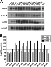
Human MAb reactivities to HeV G glycoprotein stalk mutants. (A) Alanine substitution mutants of HeV G were transiently expressed in HeLa-USU cells, metabolically labeled, and chased as described in Materials and Methods. Cell lysates were prepared and divided equally into four parts, which were immunoprecipitated with human MAbs 101, 102.4, and 106 or rabbit polyclonal G-specific antiserum (control). The precipitated proteins were analyzed by SDS-PAGE followed by autoradiography. (B) Autoradiographs were used to quantify the relative amounts of precipitated protein by spot densitometry, and the results are expressed as the percentage of WT reactivity.

References

    1. Aguilar, H. C., K. A. Matreyek, C. M. Filone, S. T. Hashimi, E. L. Levroney, O. A. Negrete, A. Bertolotti-Ciarlet, D. Y. Choi, I. McHardy, J. A. Fulcher, S. V. Su, M. C. Wolf, L. Kohatsu, L. G. Baum, and B. Lee. 2006. N-glycans on Nipah virus fusion protein protect against neutralization but reduce membrane fusion and viral entry. J. Virol. 804878-4889. - PMC - PubMed
    1. Anonymous. 2004. Hendra virus—Australia (Queensland) ProMED archive no. 20041214.3307. International Society for Infectious Diseases.
    1. Anonymous. 2008. Hendra virus, human, equine—Australia (04): (Queensland) ProMED archive no. 20080725.2260. International Society for Infectious Diseases.
    1. Anonymous. 2004. Person-to-person transmission of Nipah virus during outbreak in Faridpur District, 2004. Health Sci. Bull. 25-9.
    1. Bishop, K. A., T. S. Stantchev, A. C. Hickey, D. Khetawat, K. N. Bossart, V. Krasnoperov, P. Gill, Y. R. Feng, L. Wang, B. T. Eaton, L. F. Wang, and C. C. Broder. 2007. Identification of Hendra virus G glycoprotein residues that are critical for receptor binding. J. Virol. 815893-5901. - PMC - PubMed

LinkOut - more resources