

Retroviruses human immunodeficiency virus and murine leukemia virus are enriched in phosphoinositides
- PMID: 18799574
- PMCID: PMC2573248
- DOI: 10.1128/JVI.00981-08
Retroviruses human immunodeficiency virus and murine leukemia virus are enriched in phosphoinositides
Abstract
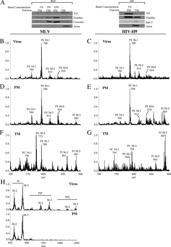
Retroviruses acquire a lipid envelope during budding from the membrane of their hosts. Therefore, the composition of this envelope can provide important information about the budding process and its location. Here, we present mass spectrometry analysis of the lipid content of human immunodeficiency virus type 1 (HIV-1) and murine leukemia virus (MLV). The results of this comprehensive survey found that the overall lipid content of these viruses mostly matched that of the plasma membrane, which was considerably different from the total lipid content of the cells. However, several lipids are enriched in comparison to the composition of the plasma membrane: (i) cholesterol, ceramide, and GM3; and (ii) phosphoinositides, phosphorylated derivatives of phosphatidylinositol. Interestingly, microvesicles, which are similar in size to viruses and are also released from the cell periphery, lack phosphoinositides, suggesting a different budding mechanism/location for these particles than for retroviruses. One phosphoinositide, phosphatidylinositol 4,5-bisphosphate [PI(4,5)P(2)], has been implicated in membrane binding by HIV Gag. Consistent with this observation, we found that PI(4,5)P(2) was enriched in HIV-1 and that depleting this molecule in cells reduced HIV-1 budding. Analysis of mutant virions mapped the enrichment of PI(4,5)P(2) to the matrix domain of HIV Gag. Overall, these results suggest that HIV-1 and other retroviruses bud from cholesterol-rich regions of the plasma membrane and exploit matrix/PI(4,5)P(2) interactions for particle release from cells.
Figures


Similar articles
-
Quantification of phosphoinositides reveals strong enrichment of PIP2 in HIV-1 compared to producer cell membranes.Sci Rep. 2019 Nov 27;9(1):17661. doi: 10.1038/s41598-019-53939-z. Sci Rep. 2019. PMID: 31776383 Free PMC article.
-
Elucidating the mechanism by which compensatory mutations rescue an HIV-1 matrix mutant defective for gag membrane targeting and envelope glycoprotein incorporation.J Mol Biol. 2015 Mar 27;427(6 Pt B):1413-1427. doi: 10.1016/j.jmb.2015.01.018. Epub 2015 Feb 7. J Mol Biol. 2015. PMID: 25659909 Free PMC article.
-
Multiple Gag domains contribute to selective recruitment of murine leukemia virus (MLV) Env to MLV virions.J Virol. 2013 Feb;87(3):1518-27. doi: 10.1128/JVI.02604-12. Epub 2012 Nov 14. J Virol. 2013. PMID: 23152533 Free PMC article.
-
The Interplay between HIV-1 Gag Binding to the Plasma Membrane and Env Incorporation.Viruses. 2020 May 16;12(5):548. doi: 10.3390/v12050548. Viruses. 2020. PMID: 32429351 Free PMC article. Review.
-
Roles played by acidic lipids in HIV-1 Gag membrane binding.Virus Res. 2014 Nov 26;193:108-15. doi: 10.1016/j.virusres.2014.06.015. Epub 2014 Jul 3. Virus Res. 2014. PMID: 24998886 Free PMC article. Review.
Cited by
-
Retroviral env glycoprotein trafficking and incorporation into virions.Mol Biol Int. 2012;2012:682850. doi: 10.1155/2012/682850. Epub 2012 Jul 2. Mol Biol Int. 2012. PMID: 22811910 Free PMC article.
-
Targeting of murine leukemia virus gag to the plasma membrane is mediated by PI(4,5)P2/PS and a polybasic region in the matrix.J Virol. 2010 Jan;84(1):503-15. doi: 10.1128/JVI.01134-09. J Virol. 2010. PMID: 19828619 Free PMC article.
-
Opposing mechanisms involving RNA and lipids regulate HIV-1 Gag membrane binding through the highly basic region of the matrix domain.Proc Natl Acad Sci U S A. 2010 Jan 26;107(4):1600-5. doi: 10.1073/pnas.0908661107. Epub 2010 Jan 4. Proc Natl Acad Sci U S A. 2010. PMID: 20080620 Free PMC article.
-
Phospholipase D functional ablation has a protective effect in an Alzheimer's disease Caenorhabditis elegans model.Sci Rep. 2018 Feb 23;8(1):3540. doi: 10.1038/s41598-018-21918-5. Sci Rep. 2018. PMID: 29476137 Free PMC article.
-
Cholesterol-dependent membrane fusion induced by the gp41 membrane-proximal external region-transmembrane domain connection suggests a mechanism for broad HIV-1 neutralization.J Virol. 2014 Nov;88(22):13367-77. doi: 10.1128/JVI.02151-14. Epub 2014 Sep 10. J Virol. 2014. PMID: 25210180 Free PMC article.
References
Publication types
MeSH terms
Substances
Grants and funding
LinkOut - more resources
Full Text Sources
Other Literature Sources
Research Materials
Miscellaneous
