

Mutational analysis of vaccinia virus mRNA cap (guanine-N7) methyltransferase reveals essential contributions of the N-terminal peptide that closes over the active site
- PMID: 18799596
- PMCID: PMC2578867
- DOI: 10.1261/rna.1201308
Mutational analysis of vaccinia virus mRNA cap (guanine-N7) methyltransferase reveals essential contributions of the N-terminal peptide that closes over the active site
Abstract
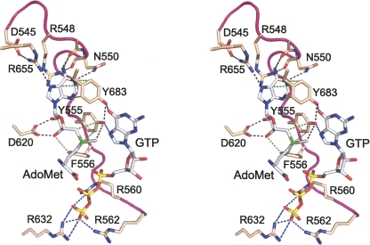
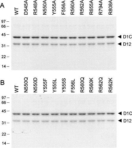
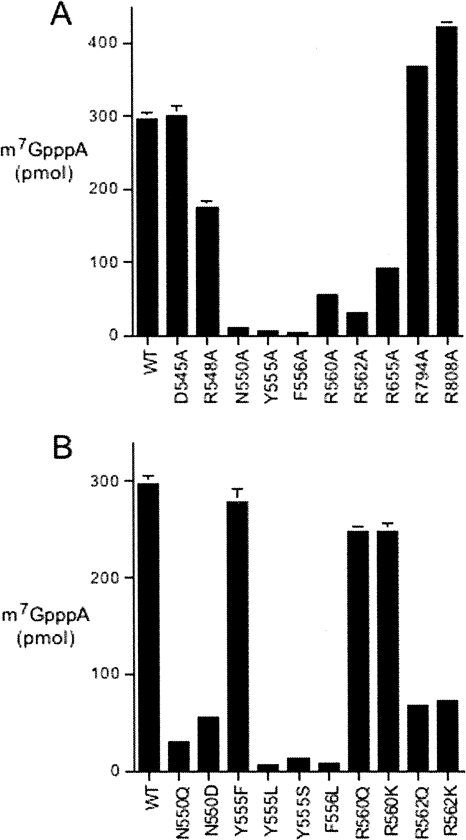
RNA guanine-N7 methyltransferase catalyzes the third step of eukaryal mRNA capping, the transfer of a methyl group from AdoMet to GpppRNA to form m(7)GpppRNA. Mutational and crystallographic analyses of cellular and poxvirus cap methyltransferases have yielded a coherent picture of a conserved active site and determinants of substrate specificity. Models of the Michaelis complex suggest a direct in-line mechanism of methyl transfer. Because no protein contacts to the guanine-N7 nucleophile, the AdoMet methyl carbon (Cepsilon) or the AdoHcy sulfur (Sdelta) leaving group were observed in ligand-bound structures of cellular cap methyltransferase, it was initially thought that the enzyme facilitates catalysis by optimizing proximity and geometry of the donor and acceptor. However, the structure of AdoHcy-bound vaccinia virus cap methyltransferase revealed the presence of an N-terminal "lid peptide" that closes over the active site and makes multiple contacts with the substrates, including the AdoMet sulfonium. This segment is disordered in the vaccinia apoenzyme and is not visible in the available structures of cellular cap methyltransferase. Here, we conducted a mutational analysis of the vaccinia virus lid peptide ((545)DKFRLNPEVSYFTNKRTRG(563)) entailing in vivo and in vitro readouts of the effects of alanine and conservative substitutions. We thereby identified essential functional groups that interact with the AdoMet sulfonium (Tyr555, Phe556), the AdoMet adenine (Asn550), and the cap triphosphate bridge (Arg560, Arg562). The results suggest that van der Waals contacts of Tyr555 and Phe556 to the AdoMet Sdelta and C epsilon atoms, and the electron-rich environment around the sulfonium, serve to stabilize the transition state of the transmethylation reaction.
Figures
Similar articles
-
Genetic analysis of poxvirus mRNA cap methyltransferase: suppression of conditional mutations in the stimulatory D12 subunit by second-site mutations in the catalytic D1 subunit.Virology. 2006 Aug 15;352(1):145-56. doi: 10.1016/j.virol.2006.03.050. Epub 2006 May 23. Virology. 2006. PMID: 16716374
-
Structure and mechanism of mRNA cap (guanine-N7) methyltransferase.Mol Cell. 2004 Jan 16;13(1):77-89. doi: 10.1016/s1097-2765(03)00522-7. Mol Cell. 2004. PMID: 14731396
-
Vaccinia virus mRNA (guanine-7-)methyltransferase: mutational effects on cap methylation and AdoHcy-dependent photo-cross-linking of the cap to the methyl acceptor site.Biochemistry. 1996 May 28;35(21):6900-10. doi: 10.1021/bi960221a. Biochemistry. 1996. PMID: 8639642
-
Structure-function analysis of vaccinia virus mRNA cap (guanine-N7) methyltransferase.RNA. 2008 Apr;14(4):696-705. doi: 10.1261/rna.928208. Epub 2008 Feb 6. RNA. 2008. PMID: 18256245 Free PMC article.
-
Structure-function analysis of the triphosphatase component of vaccinia virus mRNA capping enzyme.J Virol. 1997 Dec;71(12):9837-43. doi: 10.1128/JVI.71.12.9837-9843.1997. J Virol. 1997. PMID: 9371657 Free PMC article.
Cited by
-
Structure-function analysis of severe acute respiratory syndrome coronavirus RNA cap guanine-N7-methyltransferase.J Virol. 2013 Jun;87(11):6296-305. doi: 10.1128/JVI.00061-13. Epub 2013 Mar 27. J Virol. 2013. PMID: 23536667 Free PMC article.
-
Burkholderia glumae ToxA Is a Dual-Specificity Methyltransferase That Catalyzes the Last Two Steps of Toxoflavin Biosynthesis.Biochemistry. 2016 May 17;55(19):2748-59. doi: 10.1021/acs.biochem.6b00167. Epub 2016 May 3. Biochemistry. 2016. PMID: 27070241 Free PMC article.
-
Crystal structure of vaccinia virus mRNA capping enzyme provides insights into the mechanism and evolution of the capping apparatus.Structure. 2014 Mar 4;22(3):452-65. doi: 10.1016/j.str.2013.12.014. Structure. 2014. PMID: 24607143 Free PMC article.
-
Identification of aromatic amino acid residues in conserved region VI of the large polymerase of vesicular stomatitis virus is essential for both guanine-N-7 and ribose 2'-O methyltransferases.Virology. 2010 Dec 20;408(2):241-52. doi: 10.1016/j.virol.2010.09.017. Epub 2010 Oct 18. Virology. 2010. PMID: 20961592 Free PMC article.
-
Enzymology of RNA cap synthesis.Wiley Interdiscip Rev RNA. 2010 Jul-Aug;1(1):152-72. doi: 10.1002/wrna.19. Epub 2010 May 25. Wiley Interdiscip Rev RNA. 2010. PMID: 21956912 Free PMC article. Review.
References
-
- Bujnicki J.M., Feder M., Radlinska M., Rychlewski L. mRNA:guanine-N7 methyltransferases: Identification of novel members of the family, evolutionary analysis, homology modeling, and analysis of sequence-structure-function relationships. BMC Bioinformatics. 2001;2:2. doi: 10.1186/1471-2105-2-2. - DOI - PMC - PubMed
-
- Cong P., Shuman S. Methyltransferase and subunit association domains of vaccinia virus mRNA capping enzyme. J. Biol. Chem. 1992;267:16424–16429. - PubMed
-
- Fabrega C., Hausmann S., Shen V., Shuman S., Lima C.D. Structure and mechanism of cap (guanine-N7) methyltransferase. Mol. Cell. 2004;13:77–89. - PubMed
-
- Hausmann S., Zheng S., Fabrega C., Schneller S.W., Lima C.D., Shuman S. Encephalitozoon cuniculi mRNA cap (guanine-N7) methyltransferase: Methyl acceptor specificity, inhibition by AdoMet analogs, and structure-guided mutational analysis. J. Biol. Chem. 2005;280:20404–20412. - PubMed
Publication types
MeSH terms
Substances
Grants and funding
LinkOut - more resources
Full Text Sources
Other Literature Sources
Miscellaneous