. 2008 Nov;19(11):5006-18.
doi: 10.1091/mbc.e08-01-0034. Epub 2008 Sep 17.

Retrograde flow and myosin II activity within the leading cell edge deliver F-actin to the lamella to seed the formation of graded polarity actomyosin II filament bundles in migrating fibroblasts

Affiliations

Retrograde flow and myosin II activity within the leading cell edge deliver F-actin to the lamella to seed the formation of graded polarity actomyosin II filament bundles in migrating fibroblasts

Tom W Anderson et al. Mol Biol Cell. 2008 Nov.

Abstract

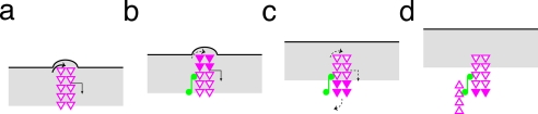
In migrating fibroblasts actomyosin II bundles are graded polarity (GP) bundles, a distinct organization to stress fibers. GP bundles are important for powering cell migration, yet have an unknown mechanism of formation. Electron microscopy and the fate of photobleached marks show actin filaments undergoing retrograde flow in filopodia, and the lamellipodium are structurally and dynamically linked with stationary GP bundles within the lamella. An individual filopodium initially protrudes, but then becomes separated from the tip of the lamellipodium and seeds the formation of a new GP bundle within the lamella. In individual live cells expressing both GFP-myosin II and RFP-actin, myosin II puncta localize to the base of an individual filopodium an average 28 s before the filopodium seeds the formation of a new GP bundle. Associated myosin II is stationary with respect to the substratum in new GP bundles. Inhibition of myosin II motor activity in live cells blocks appearance of new GP bundles in the lamella, without inhibition of cell protrusion in the same timescale. We conclude retrograde F-actin flow and myosin II activity within the leading cell edge delivers F-actin to the lamella to seed the formation of new GP bundles.


Figures

Figure 1.
Figure 1.
Actin filaments within the leading cell edge and GP bundles are structurally contiguous. (a and b) A migrating chick embryo fibroblast fixed and stained for F-actin with Alexa 594-phalloidin. (a) Labels indicate the lamellipodium (lp), lamella (la), nuclear region (n), and cell rear (r). A dashed line indicates the boundary between the lamellipodium and lamella. Solid arrowheads indicate GP bundles. (b) An enlarged view of the box region in panel a shows the region of the lamellipodium and the front part of the lamella in more detail. Hollow arrowheads indicate filopodia embedded within the lamellipodium, and solid arrowheads indicate GP actin filament bundles at the front of the lamella. (c–g) Whole mount EM images (c–f) of the front of the cell in two (c, e and d, f) migrating fibroblasts. The GP bundle in the boxed region (c and d) is located at the lamellipodium–lamella boundary. (e and f) Enlarged views of the boxed regions in c and d, respectively; the boundary between the lamellipodium and lamella is indicated with a dashed red line, and an actin filament from the tip of a GP bundle in the lamella (lower red arrowhead) is contiguous with the actin filament (upper red arrowhead) within a filopodium (e) or lamellipodium (f). (g) Quantification of tracing the spatial location of actin filaments within the ends of GP bundles visualized in EM images (average, n = 230 GP bundle ends in seven cells). Lp denotes contiguous with F-actin within the lamellipodium, and fp denotes contiguous with F-actin within a filopodium. Variation in appearance of the two different cells in c and d is due to slight difference in the critical point drying. Apparent actin filament width is increased due to platinum and carbon coating (see Materials and Methods). Bars, (a) 18 μm, (b) 3 μm, (c and d) 1 μm, and (e and f) 100 nm.
Figure 2.
Figure 2.
Actin filaments within the leading cell edge and lamella are dynamically linked. Live cells expressing GFP-actin were imaged every 1 s with a SP5 confocal microscope, and a subzone within the leading cell edge was photobleached and then tracked. (a–e) Retrograde F-actin flow (rate in this lamellipodium is 0.64 um/min) with respect to the substratum occurs through the lamellipodium (b and c, dark band moves leftwards in the box), reaches the boundary with the lamella (c and d), and stops flowing within the lamella (d and e, no movement leftwards at the rear of the mark). (f) Kymograph (4 μm across) of the box region in a–e. Diagonal dashed line shows retrograde F-actin flow within the lamellipodium, and vertical dashed line shows no flow within the lamella. Gray scale value intensities were colored with a cool hue look up table in ImageJ (http://rsb.info.nih.gov/ij/). The photobleached zone appears purple/blue. (g) Quantification of behavior of the zone originally bleached within the lamellipodium and tracked to the lamella (n = 35 bleached zones in 19 live cells). (h–l) Leading cell edge and lamella of a different cell: a filopodium is indicated with an arrow (h), a zone on the filopodium is photobleached (i) and tracked (i–k), and the bleached zone is then visible within a GP bundle in the lamella (k) that recovers by panel l. Bars, (e and l) 5 μm.
Figure 3.
Figure 3.
F-actin within filopodia and lamellipodia seed the formation of new GP actin filament bundles within the lamella. The origin of individual GP bundles in live cells expressing GFP-actin was determined from their time-lapse prehistories imaged by spinning disk confocal (a–f and i) or confocal (g and h) microscopy. (a) Quantification of the origin of GP bundles classified as filopodia (fp) or subzones within the lamellipodium (lp) at 10-s temporal resolution (n = 50 events in 10 live cells). (b) An individual cell's migration over 20 min. (c) An enlargement of the boxed region in panel b, showing an individual example of a filopodium (hollow arrowhead, −50 to −10 s) that seeds the formation (0 s) of a nascent GP bundle (solid arrowhead, 0–100 s) and subsequent GP bundle elongation (compare guillemets, 0–100 s). (d) Three individual examples of filopodium-to-GP bundle conversion events (solid magenta line is quantification of the filopodium-to-GP bundle conversion in panel c. (e) Quantification of filopodium protrusion rate (average 1.39 ± 0.15 μm/min) and GP bundle movement at tip (average −0.05 ± 0.005 μm/min; n = 50 individual pairs of filopodium-to-GP bundle conversion events in 10 live cells). (f) Three individual examples of nascent GP bundle elongation (solid magenta line represents the nascent GP bundle imaged in panel c). (g) Higher time resolution (imaged every 1 s) of a filopodium-to-GP bundle conversion event. Arrow either indicates the filopodium (−5 s) or tip of the leading cell edge (0, 6, 12, 13, and 18 s). Conversion of a filopodium to a GP bundle (0 s) is defined as a clear gap between the tip of the leading cell edge (0 s, arrow) and the tip of the filopodium (0 s, arrowhead). Arrowheads indicate the tip of the F-actin bundle and guillemets indicate the rear of the bundle. (h) Quantification of the origin of GP bundles classified as filopodia (fp) or subzones within the lamellipodium (lp) at 1-s temporal resolution (n = 57 events in 11 live cells). (i) Example of a filopodium-to-GP bundle conversion with minimal bundle shortening. Bars, (b) 18 μm, (c, i, and g) 2 μm.
Figure 4.
Figure 4.
Nascent GP bundles within the lamella elongate by subunit assembly in the front to rearwards direction. A live cell expressing GFP-actin was imaged every 1 s with a SP5 confocal microscope, and a subzone within several adjacent nascent GP bundles within the front of the lamella (one of bundles illustrated in a, between arrows) was photobleached (b, between arrows; long arrow indicates the front of the mark and short arrow indicates the rear of the mark). Recovery of fluorescence is in the front-to-rearward direction (b–d, long arrow moves to short arrow). Note, in contrast to the behavior of marks when localized in the lamellipodium (Figure 2, a–c and f) the position of rear of mark in the GP bundle (short arrow) does not move. (e) Kymograph analysis (region analyzed shown in boxed region in f) illustrating fluorescence recovery; rear of mark does not move (horizontal dashed line), whereas recovery occurs from front-to-rearward direction (diagonal dashed-line). (g) Quantification of fluorescence recovery (n = 54 bundles in 17 live cells). (a–d) Front and F indicate from front-to-rearward direction; B indicates from back-to-frontward direction. Bar, (d) 5 μm.
Figure 5.
Figure 5.
Myosin II puncta localize to the leading cell edge. Cell expressing GFP-myosin II light chain (b and e) fixed and costained for F-actin with Alexa 594-phalloidin (a and d). (d and e) Enlarged view of the box in a and b, respectively. (c and f) Overlay images. Note, myosin II puncta (e, arrowheads) are localized within the leading cell edge (below dashed line) and at the rear of the leading cell edge at the boundary with the lamella (dashed line). (g) Quantification of cells with myosin II puncta detected in the leading cell edge (le). (h) Quantification of position of myosin II puncta detected in the leading cell edge (le). (i) Quantification of myosin II puncta associated with F-actin intensity in the leading cell edge (le) (for panels g–i, n = 127 puncta in 19 cells). Bar, (a) 10 μm, (d) 1 μm.
Figure 6.
Figure 6.
Myosin II localizes to filopodia destined to form GP actin filament bundles. Live cell expressing both RFP-actin (a, top panels) and GFP-myosin II light chain (a, middle panels) was imaged by dual-color time-lapse spinning disk confocal microscopy (both F-actin in red and myosin II in green are traced in the bottom panels). Conversion of a filopodium (hollow arrowheads) to a GP bundle (solid arrowheads) is illustrated. Time (sec) is relative to conversion (0 s). (b) Quantification of initial spatial location of detected myosin II puncta destined to associate with a nascent GP bundle (n = 20 events in four live cells). Boundary indicates boundary of the leading cell edge and lamella. (c) Quantification of spatial position of myosin II associated with F-actin destined to convert to a GP bundle. (d) Quantification of relative temporal association of myosin II with a filopodium destined to form a GP bundle (for c and d, n = 10 conversion events in two live cells). Bar, (a) 2 μm.
Figure 7.
Figure 7.
Myosin II ATPase activity is required for appearance of new GP bundles in the lamella. Live cell infected and expressing RFP-actin only and treated with 100 μM (±)-blebbistatin to block myosin II activity; time in panel a indicates minutes relative to addition of the inhibitor. Boxes in panel a indicate the area of greatest protrusion over the preceding 10 min. (b) Enlargements of the boxed areas in panel a; arrowheads indicate GP bundles. Note the absence of new GP bundles in the lamella during 10-min blebbistatin treatment (bottom panel). (c) Quantification of the entire front of the lamella of the density of GP actin bundles before (an average 0.055 ± 0.007 per μm2) and after (an average 0.006 ± 0.004 per μm2) addition of 100 μM (±)-blebbistatin for 10 min in the same live cell (n = eight live cells). Bars, (a) 20 μm, (b) 5 μm.
Figure 8.
Figure 8.
Retrograde flow delivers F-actin to the lamella to seed the formation of GP bundles. A simplified schematic based on data on F-actin and myosin II dynamics and actin filament polarity from our current and previous work in migrating fibroblasts and other cited information (see text for details). Observed behavior of marked F-actin (closed magenta chevrons) in the lamellipodium (gray stripe) and lamella (white region) is represented. (a, protrusion) local F-actin in a filopodium or subzone within the lamellipodium (magenta chevrons) undergoing retrograde flow (bent arrow) assembles at its tip (curly-on arrow) at the same rate as bulk F-actin within the leading cell edge (gray stripe). Not shown, for simplicity, is F-actin disassembly, which also occurs within the F-actin network. (b, association of myosin II) A myosin II punctum (green dumb-bell) associates with the base of the local F-actin region (magenta chevrons) destined to form a GP bundle. (b and c, conversion) Local F-actin continues to flow retrograde (bent arrow) as local assembly rate slows down (dashed curly-on arrow), whereas net cell protrusion (black line) is driven at the prior assembly rate. This results in separation (gap in c) of the tip of the local F-actin region from the tip of the leading cell edge. (c and d, F-actin seed) This process delivers an F-actin seed (magenta chevrons) to the boundary (interface of white and gray regions) of the leading cell edge (gray stripe) and the lamella (white region). Detectable retrograde F-actin flow slows down/stops (dashed bent arrow) in the seed through activation of cell–substratum adhesion formation. (d, GP bundle elongation) Associated myosin II (green dumb-bells) promotes recruitment of oppositely opposed actin filaments, as required for graded polarity in this region. Actin subunits during elongation (compare c and d, open chevrons) mainly assemble at filament barbed-ends in the front-to-rear direction. Our data predicts either balanced local actin disassembly in the original F-actin seed at conversion to a GP bundle (c, dashed curly-off arrow) and/or GP bundle elongation is driven primarily by addition of subunits recruited to associated myosin II (c and d, compare open chevrons).

References

    1. Badley R. A., Couchman J. R., Rees D. A. Comparison of the cell cytoskeleton in migratory and stationary chick fibroblasts. J. Muscle Res. Cell Motil. 1980;1:5–14. - PubMed
    1. Bernstein B. W., Bamburg J. R. A proposed mechanism for cell polarization with no external cues. Cell Motil. Cytoskelet. 2004;58:96–103. - PubMed
    1. Bi E., Maddox P., Lew D. J., Salmon E. D., McMillan J. N., Yeh E., Pringle J. R. Involvement of an actomyosin contractile ring in Saccharomyces cerevisiae cytokinesis. J. Cell Biol. 1998;142:1301–1312. - PMC - PubMed
    1. Burgess D. R. Cytokinesis: new roles for myosin. Curr. Biol. 2005;15:R310–R311. - PubMed
    1. Carlier M. F., Pantaloni D. Control of actin assembly dynamics in cell motility. J. Biol. Chem. 2007;282:23005–23009. - PubMed

Publication types

LinkOut - more resources