. 2008 Nov;295(5):C1376-84.
doi: 10.1152/ajpcell.00362.2008. Epub 2008 Sep 17.

Type 1 inositol 1,4,5-trisphosphate receptors mediate UTP-induced cation currents, Ca2+ signals, and vasoconstriction in cerebral arteries

Affiliations

Type 1 inositol 1,4,5-trisphosphate receptors mediate UTP-induced cation currents, Ca2+ signals, and vasoconstriction in cerebral arteries

Guiling Zhao et al. Am J Physiol Cell Physiol. 2008 Nov.

Erratum in

  • Am J Physiol Cell Physiol. 2009 Jul;297(1):C226

Abstract

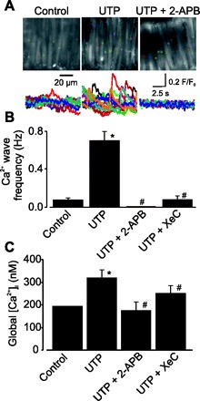
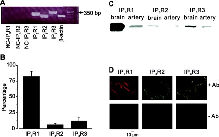
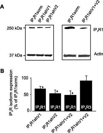
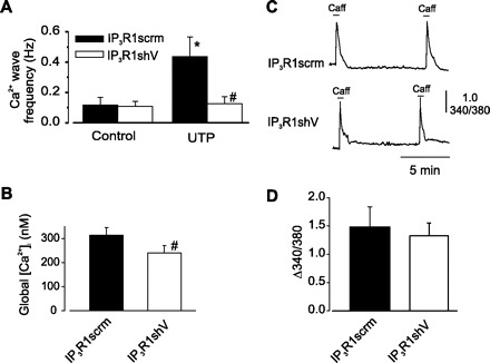
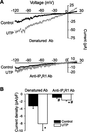
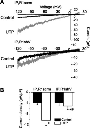
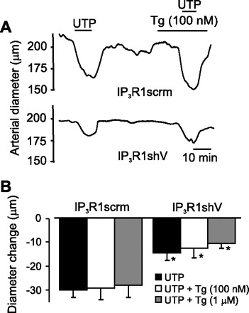
Inositol 1,4,5-trisphosphate receptors (IP(3)Rs) regulate diverse physiological functions, including contraction and proliferation. There are three IP(3)R isoforms, but their functional significance in arterial smooth muscle cells is unclear. Here, we investigated relative expression and physiological functions of IP(3)R isoforms in cerebral artery smooth muscle cells. We show that 2-aminoethoxydiphenyl borate and xestospongin C, membrane-permeant IP(3)R blockers, reduced Ca(2+) wave activation and global intracellular Ca(2+) ([Ca(2+)](i)) elevation stimulated by UTP, a phospholipase C-coupled purinergic receptor agonist. Quantitative PCR, Western blotting, and immunofluorescence indicated that all three IP(3)R isoforms were expressed in acutely isolated cerebral artery smooth muscle cells, with IP(3)R1 being the most abundant isoform at 82% of total IP(3)R message. IP(3)R1 knockdown with short hairpin RNA (shRNA) did not alter baseline Ca(2+) wave frequency and global [Ca(2+)](i) but abolished UTP-induced Ca(2+) wave activation and reduced the UTP-induced global [Ca(2+)](i) elevation by approximately 61%. Antibodies targeting IP(3)R1 and IP(3)R1 knockdown reduced UTP-induced nonselective cation current (I(cat)) activation. IP(3)R1 knockdown also reduced UTP-induced vasoconstriction in pressurized arteries with both intact and depleted sarcoplasmic reticulum (SR) Ca(2+) by approximately 45%. These data indicate that IP(3)R1 is the predominant IP(3)R isoform expressed in rat cerebral artery smooth muscle cells. IP(3)R1 stimulation contributes to UTP-induced I(cat) activation, Ca(2+) wave generation, global [Ca(2+)](i) elevation, and vasoconstriction. In addition, IP(3)R1 activation constricts cerebral arteries in the absence of SR Ca(2+) release by stimulating plasma membrane I(cat).


Figures

Fig. 1.
Fig. 1.
UTP activates Ca2+ waves and elevates global intracellular Ca2+ concentration ([Ca2+]i) as a result of inositol 1,4,5-trisphosphate (IP3) receptor (IP3R) activation in cerebral artery smooth muscle cells. A: Ca2+ signals recorded in smooth muscle cells of an intact cerebral artery under control conditions and in the presence of UTP (30 μM) and UTP + 2-aminoethoxydiphenyl borate (2-APB, 100 μM). Top: colored boxes (2.2 × 2.2 μm, 10 × 10 pixels) indicate locations of changes in fluorescence ratio (F/F0) measured over 10 s in arterial smooth muscle cells. Bottom: colored traces showing changes in F/F0 for respective colored boxes over 10 s. Images were acquired at 30 Hz. B: mean data illustrating inhibition of UTP-induced Ca2+ waves in smooth muscle cells of cerebral artery segments by 2-APB (100 μM) and xestospongin C (XeC, 20 μM). C: UTP-induced global [Ca2+]i elevations are abolished by 2-APB and attenuated by XeC. Changes in global [Ca2+]i were calculated from a control value previously determined using fura-2 (10). Values are means ± SE; n = 27, 13, 4, and 9 for control, UTP, UTP + 2-APB, and UTP + XeC, respectively. *P < 0.05 vs. control. #P < 0.05 vs. UTP.
Fig. 2.
Fig. 2.
PCR, Western analysis, and immunofluorescence identify IP3R1, IP3R2, and IP3R3 expression in cerebral artery smooth muscle cells. A: RT-PCR detection of IP3R1 (285 bp), IP3R2 (241 bp), IP3R3 (319 bp), and β-actin (257 bp) in isolated smooth muscle cells. NC, negative control using smooth muscle cell RNA, instead of cDNA. Right: 50-bp-increment ladders are shown. B: real-time PCR data indicating percent message for IP3R1, IP3R2, and IP3R3 in isolated smooth muscle cells. Values are means ± SE; n = 3 for each. C: Western blot detection of IP3R1, IP3R2, and IP3R3 in cerebral arteries and brain. D: immunofluorescence results showing IP3R1, IP3R2, and IP3R3 expression and localization in isolated smooth muscle cells.
Fig. 3.
Fig. 3.
IP3R1 short hairpin RNA (shRNA) suppression vectors attenuate IP3R1 protein in cerebral arteries. A: Western blot illustrating that IP3R1shV1, IP3R1shV2, and IP3R1shV1 + IP3R1shV2 reduce IP3R1 protein in cerebral arteries. B: mean data illustrating that shRNAs targeting IP3R1 reduce IP3R1 expression, but do not alter IP3R3 expression, in cerebral arteries. Values are means ± SE; n = 5, 4, 3, and 3 for IP3R1shV1, IP3R1shV2, and IP3R1shV1 + IP3R1shV2 (IP3R1) and IP3R1shV1 + IP3R1shV2 (IP3R3), respectively. *P < 0.05 vs. IP3R1scrm.
Fig. 4.
Fig. 4.
IP3R1 knockdown attenuates UTP- induced Ca2+ wave generation and global [Ca2+]i elevation in smooth muscle cells of intact cerebral artery segments. A: IP3R1 knockdown did not alter resting Ca2+ wave frequency but altered UTP-induced Ca2+ wave generation (n = 6). B: IP3R1 knockdown reduced UTP-induced global [Ca2+]i elevation (n = 6). Values show mean change in calibrated global [Ca2+]i from a control value of 193 nM (10). C: amplitude of caffeine (10 mM)-induced [Ca2+]i transients were similar in IP3R1scrm- and IP3R1shV-treated cerebral arteries. D: mean change in fura-2 ratio (Δ340/380) in cerebral arteries treated with IP3R1scrm (n = 16) and IP3R1shV (n = 14). Values are means ± SE. *P < 0.05 vs. control. #P < 0.05 vs. IP3R1scrm.
Fig. 5.
Fig. 5.
Anti-IP3R1 antibody inhibits UTP-induced cation current (Icat) in rat cerebral artery smooth muscle cells. A: exemplar traces obtained using whole cell patch-clamp configuration with anti-IP3R1 (1:100 dilution) or its heat-denatured control in the pipette before or after UTP (30 μM) application. B: mean data showing the effect of anti-IP3R1 antibody on Icat. Values are means ± SE; n = 11 (denatured control Ab) and 5 (all others). *P < 0.05 vs. control. #P < 0.05 vs. denatured Ab.
Fig. 6.
Fig. 6.
IP3R1 knockdown inhibits Icat activation by UTP in cerebral artery smooth muscle cells. A: exemplar traces illustrating UTP-induced Icat activation in IP3R1scrm- and IP3R1shV-treated smooth muscle cells. B: mean current density data for IP3R1scrm- and IP3R1shV-treated cells. *P < 0.05 vs. control. #P < 0.05 vs. IP3R1scrm.
Fig. 7.
Fig. 7.
IP3R1 knockdown attenuates UTP-induced constriction in pressurized (20 mmHg) cerebral arteries. A: exemplar diameter measurements illustrating UTP-induced vasoconstriction in cerebral arteries treated with IP3R1scrm or IP3R1shV in the presence or absence of thapsigargin (Tg, 100 nM). B: mean diameter change data. Values are means ± SE; n = 6 (UTP and UTP + 100 nM Tg) and 4 (UPT + 1 μM Tg). *P < 0.05 vs. IP3R1scrm.

Similar articles

Cited by

References

    1. Acharya JK, Jalink K, Hardy RW, Hartenstein V, Zuker CS. InsP3 receptor is essential for growth and differentiation but not for vision in Drosophila Neuron 18: 881–887, 1997 - PubMed
    1. Albert AP, Large WA. Store-operated Ca2+-permeable non-selective cation channels in smooth muscle cells. Cell Calcium 33: 345–356, 2003 - PubMed
    1. Albert AP, Large WA. Synergism between inositol phosphates and diacylglycerol on native TRPC6-like channels in rabbit portal vein myocytes. J Physiol 552: 789–795, 2003 - PMC - PubMed
    1. Albert AP, Large WA. Inhibitory regulation of constitutive transient receptor potential-like cation channels in rabbit ear artery myocytes. J Physiol 560: 169–180, 2004 - PMC - PubMed
    1. Blatter LA, Wier WG. Agonist-induced [Ca2+]i waves and Ca2+-induced Ca2+ release in mammalian vascular smooth muscle cells. Am J Physiol Heart Circ Physiol 263: H576–H586, 1992 - PubMed

Publication types

MeSH terms