. 2008 Sep 17;28(38):9363-76.
doi: 10.1523/JNEUROSCI.1447-08.2008.

Requirement of myeloid cells for axon regeneration

Affiliations

Requirement of myeloid cells for axon regeneration

Benoit Barrette et al. J Neurosci. .

Abstract

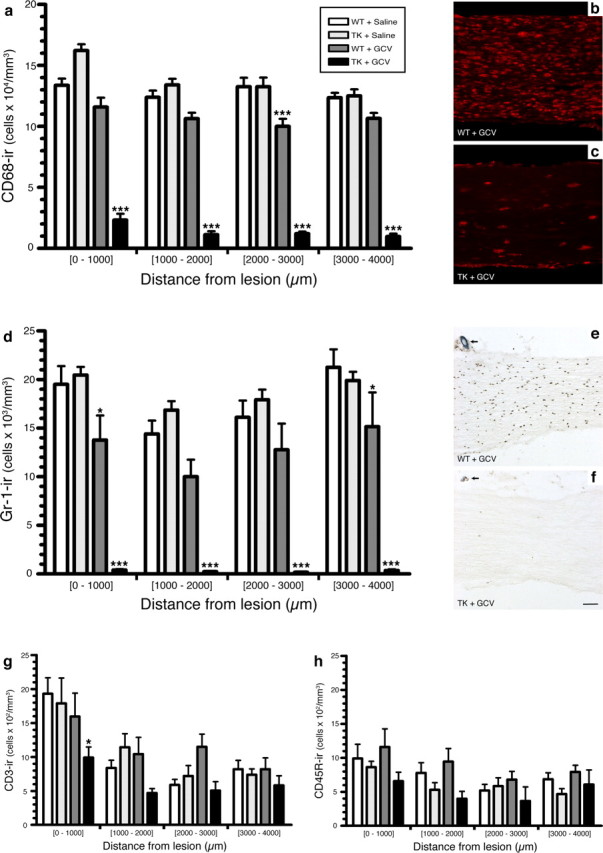
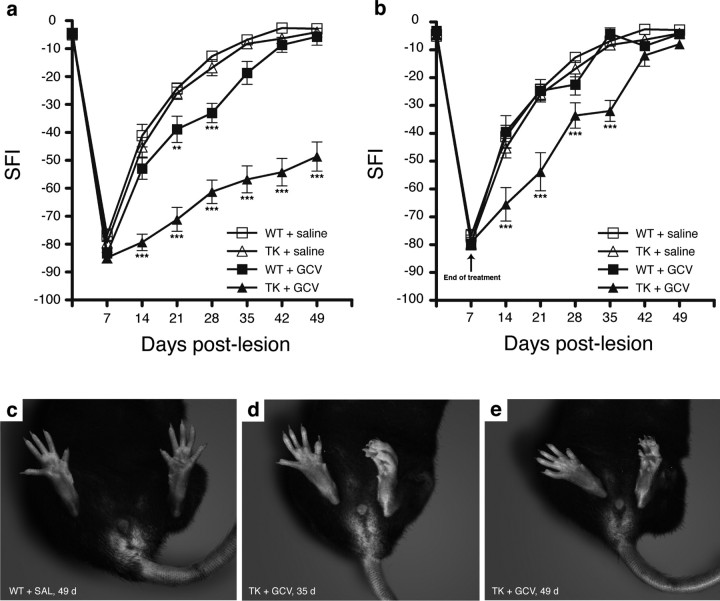
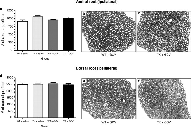
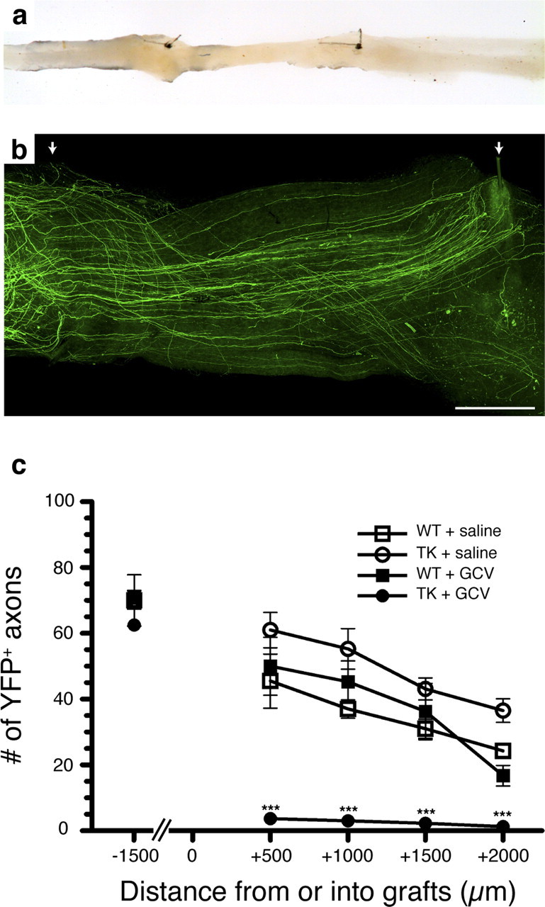
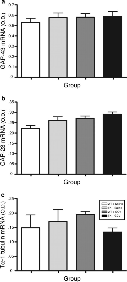
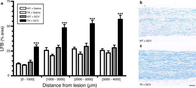
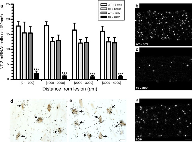
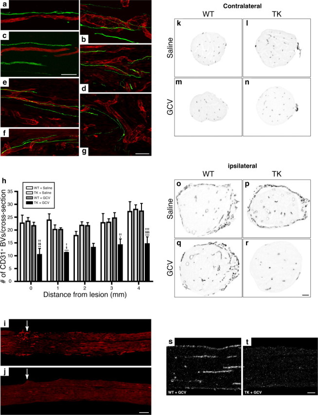
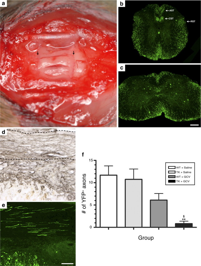
The role of CD11b+ myeloid cells in axonal regeneration was assessed using axonal injury models and CD11b-TK(mt-30) mice expressing a mutated HSV-1 thymidine kinase (TK) gene regulated by the myeloid-specific CD11b promoter. Continuous delivery of ganciclovir at a sciatic nerve lesion site greatly decreased the number of granulocytes/inflammatory monocytes and macrophages in the distal stump of CD11b-TK(mt-30) mice. Axonal regeneration and locomotor function recovery were severely compromised in ganciclovir-treated CD11b-TK(mt-30) mice. This was caused by an unsuitable growth environment rather than an altered regeneration capacity of neurons. In absence of CD11b+ cells, the clearance of inhibitory myelin debris was prevented, neurotrophin synthesis was abolished, and blood vessel formation/maintenance was severely compromised in the sciatic nerve distal stump. Spinal cord-injured axons also failed to regenerate through peripheral nerve grafts in the absence of CD11b+ cells. Therefore, myeloid cells support axonal regeneration and functional recovery by creating a growth-permissive milieu for injured axons.


Figures

Figure 1.
Figure 1.
Treatment of CD11b-TKmt-30 (TK) transgenic mice with GCV results in the almost complete depletion of CD68+ macrophages and Gr-1+ granulocytes/inflammatory monocytes in the injured sciatic nerve. a, d, g, h, Quantification of the number of CD68-immunoreactive (ir) macrophages, Gr-1-ir granulocytes/inflammatory monocytes, CD3-ir T-cells, and CD45R-ir B-cells in the sciatic nerve distal stump of TK and WT mice at 7 d after lesion in response to either GCV or saline treatment (n = 8 per group). Notably, a 89 and 98% reduction in the number of CD68+ macrophages and Gr-1+ granulocytes/inflammatory monocytes, respectively, was observed in the nerve distal stump of GCV-treated TK mice compared with saline-treated TK and WT mice at 7 d after lesion. b, c, e, f, Representative photomicrographs showing CD68 (b, c) and Gr-1 (e, f) immunostaining in the sciatic nerve distal stump of WT and TK mice treated with GCV at 7 d after lesion. The arrows point to the 10-0 suture node indicating the site of lesion (e, f). All data are expressed as mean ± SEM. ***p < 0.001 and *p < 0.05 compared with respective control groups. Scale bar: (in f) b, c, e, f, 80 μm.
Figure 2.
Figure 2.
CD11b+ cells are required for recovery of sciatic nerve functions after injury. a, Recovery of locomotor functions from a sciatic nerve lesion, as determined by the sciatic function index (SFI), is severely compromised in GCV-treated CD11b-TKmt-30 (TK) transgenic mice compared with control groups, with large deficits persisting even at 49 d after lesion (n = 12 per group). b, When minipumps were removed at 7 d after lesion in TK mice, transgenic animals recovered after 6–7 weeks (n = 12 per group). Error bars indicate SEM. c–e, Representative examples of the typical posture adopted by saline-treated WT (c) and GCV-treated TK (d, e) mice at 35 (d) and 49 (c, e) days after lesion of their left sciatic nerve. ***p < 0.001 and **p < 0.01 compared with respective control groups.
Figure 3.
Figure 3.
Depletion of CD11b+ cells does not lead to death of axotomized neurons. a, d, Counts of axonal profiles in Epon-embedded L5 ventral and dorsal roots at 50 d after lesion revealed that differences in functional recovery in CD11b-TKmt-30 (TK) transgenic mice compared with WT are not caused by the loss of motor or sensory neurons. Error bars indicate SEM. b, c, e, f, Photomicrographs showing toluidine blue staining of L5 ventral (b, c) and dorsal (e, f) roots ipsilateral to the lesion. Representative cross sections were taken from GCV-treated WT and TK mice. Scale bar: (in f) b, c, e, f, 12.5 μm.
Figure 4.
Figure 4.
Regeneration of peripheral axons is prevented in the absence of CD11b+ cells. a, Photograph showing an example of sciatic–sciatic nerve graft. b, Representative fluorescence photomicrograph showing sciatic nerve axons expressing the YFP marker that have regenerated into a PN graft collected from a saline-treated YFP/TK+ mouse (i.e., CD11b-TKmt-30 +/−). The arrows point to the two 10-0 sutures used to connect the PN graft between the proximal and distal ends of the recipient sciatic nerve from the YFP transgenic mouse. c, Quantification of the number of YFP-labeled axons at predetermined distances from the proximal host–graft interface (n = 8 per group). Although the total number of YFP+ axons was similar in all groups at a distance of 1.5 mm from the proximal host–graft interface (i.e., into the proximal end of the recipient sciatic nerve), YFP+ axons were only able to regenerate into predegenerated PN grafts in the presence of CD11b+ cells. Error bars indicate SEM. ***p < 0.001. Scale bar, 500 μm.
Figure 5.
Figure 5.
Cell bodies from axotomized sciatic motor neurons reacted normally to injury in the absence of CD11b+ cells by overexpressing regeneration-associated genes. a–c, Quantification of in situ hybridization signal for GAP-43 (a), CAP-23 (b), and Tα-1 tubulin (c) mRNAs [in optical density (O.D.); arbitrary units] in the ipsilateral motoneuronal cell group of Rexed's lamina IX at spinal levels L2–L5 in CD11b-TKmt-30+/− (TK) and WT mice treated with either GCV or saline at 7 d after sciatic nerve lesion (n = 8 per group). Error bars indicate SEM.
Figure 6.
Figure 6.
Inhibitory myelin debris could not be cleared in the absence of CD11b+ cells. a, Quantification of LFB staining of myelin in the sciatic nerve distal stump of CD11b-TKmt-30+/− (TK) and WT mice treated with either GCV or saline at 7 d after sciatic nerve lesion (n = 8 per group). Although myelin breakdown and formation of ovoids of degenerating myelin were apparent in the sciatic nerve distal stump of GCV-treated TK mice, quantification of LFB staining revealed that the load of myelin debris was >200% higher in mice depleted in CD11b+ cells compared with mice of control groups at 7 d after lesion. Error bars indicate SEM. b, c, Representative bright-field photomicrographs showing myelin stained with LFB in the sciatic nerve distal stump of GCV-treated WT and GCV-treated TK mice at 7 d after lesion. ***p < 0.001. Scale bar: (in b) b, c, 100 μm.
Figure 7.
Figure 7.
NT-3 synthesis in the injured peripheral nerve is prevented in the absence of CD11b+ cells. a, Quantification of the number of cells expressing NT-3 mRNA in the sciatic nerve distal stump of CD11b-TKmt-30 (TK) and WT mice at 7 d after lesion in response to either GCV or saline treatment (n = 8 per group). Error bars indicate SEM. b, c, Representative dark-field photomicrographs showing NT-3 mRNA expression in the sciatic nerve distal stump of GCV-treated WT (b) and GCV-treated TK (c) mice at 7 d after lesion. d, e, Photomicrographs of the sciatic nerve distal stump of a WT mouse showing colocalization of NT-3 mRNA with Gr-1-immunoreactive granulocytes/inflammatory monocytes. The arrows point to double-labeled cells. f, Representative dark-field photomicrograph showing the presence of numerous cells expressing NT-3 mRNA in the sciatic nerve distal stump of a scid mouse that has no functional T- and B-cells at 7 d after lesion. Together, these results indicate that a subset of CD11b+ myeloid cells expressing the cell surface protein Gr-1 are responsible for neurotrophin synthesis in the injured peripheral nerve. ***p < 0.001. Scale bars: (in e) d, e, 5 μm; (in f) b, c, f, 100 μm.
Figure 8.
Figure 8.
The formation/stabilization of blood vessels in the injured peripheral nerve is compromised after depletion of CD11b+ cells. a–g, Fluorescence photomicrographs showing examples of sciatic nerve- (a–c) and spinal cord-injured (d–g) axons (green) regenerating into peripheral nerve grafts in close association with blood vessels (CD31 immunolabeling; red). h, Quantification of the number of CD31-immunoreactive blood vessels at predetermined distances from the lesion site in the sciatic nerve distal stump of CD11b-TKmt-30 (TK) and WT mice at 7 d after lesion in response to either GCV or saline treatment (n = 8 per group). Error bars indicate SEM. i, j, Representative photomicrographs showing immunofluorescence for CD31 in the sciatic nerve distal stump of WT (i) and TK (j) mice treated with GCV at 7 d after lesion. The arrows point to the lesion site. k–r, Representative bright-field photomicrographs showing immunoreactivity for CD31 in sciatic nerve cross sections taken from contralateral (unlesioned) and ipsilateral (lesioned) nerves of WT and TK mice treated with either saline or GCV at 7 d after lesion. s, t, Representative dark-field photomicrographs showing Tie2 mRNA expression in the sciatic nerve distal stump of GCV-treated WT and GCV-treated TK mice at 7 d after lesion. Note the absence of Tie2 mRNA signal after depletion of CD11b+ cells. ***p < 0.001, **p < 0.01, and *p < 0.05 compared with the WT plus saline group; §§§ p < 0.001, §§ p < 0.01, and § p < 0.05 compared with the TK plus saline group; ††† p < 0.001, †† p < 0.01, and p < 0.05 compared with the WT plus GCV group. Scale bars: (in c) a–c, 50 μm; (in g) d–g, 50 μm; (in j) i, j, 250 μm; (in r) k–r, 80 μm; (in t) s, t, 100 μm.
Figure 9.
Figure 9.
Regeneration of SCI axons into PN grafts is prevented in the absence of CD11b+ cells. a, Photograph showing three sciatic nerve segments transplanted into the spinal cord of a T12/L1 dorsal hemisected Thy1-YFP-H+/− transgenic mouse. The arrows point to PN grafts/spinal cord tissue interfaces. b, c, YFP expression in the thoracic (b) and lumbar (c) spinal cord of a Thy1-YFP-H+/− transgenic mouse. Note the presence of fluorescence in axons traveling through most, if not all, spinal cord projection systems, including the descending corticospinal (CST) and rubrospinal (RST) tracts and the ascending sensory tract (AST). d, e, Visualization of laminin immunolabeling (d) (to visualize PN tissue) and YFP fluorescence (e) on adjacent spinal cord sections revealed that PN grafts are densely penetrated by regenerating SCI axons at 2 weeks after SCI/grafting. The dotted lines in d indicate the anatomical boundaries of a PN graft. f, Quantification of the number of YFP-labeled axons at predetermined distances from the rostrocaudal host–graft interface. Note that SCI YFP-labeled axons did not regenerate into predegenerated PN grafts lacking CD11b+ cells. Error bars indicate SEM. **p < 0.01 compared with the WT plus saline group; § p < 0.05 compared with the TK plus saline group. Scale bars: (in c) b, c, 275 μm; (in e) d, e, 100 μm.

References

    1. Auffray C, Fogg D, Garfa M, Elain G, Join-Lambert O, Kayal S, Sarnacki S, Cumano A, Lauvau G, Geissmann F. Monitoring of blood vessels and tissues by a population of monocytes with patrolling behavior. Science. 2007;317:666–670. - PubMed
    1. Bandtlow CE, Meyer M, Lindholm D, Spranger M, Heumann R, Thoenen H. Regional and cellular codistribution of interleukin 1 beta and nerve growth factor mRNA in the adult rat brain: possible relationship to the regulation of nerve growth factor synthesis. J Cell Biol. 1990;111:1701–1711. - PMC - PubMed
    1. Barrette B, Vallieres N, Dube M, Lacroix S. Expression profile of receptors for myelin-associated inhibitors of axonal regeneration in the intact and injured mouse central nervous system. Mol Cell Neurosci. 2007;34:519–538. - PubMed
    1. Boivin A, Pineau I, Barrette B, Filali M, Vallières N, Rivest S, Lacroix S. Toll-like receptor signaling is critical for Wallerian degeneration and functional recovery after peripheral nerve injury. J Neurosci. 2007;27:12565–12576. - PMC - PubMed
    1. Brück W, Huitinga I, Dijkstra CD. Liposome-mediated monocyte depletion during wallerian degeneration defines the role of hematogenous phagocytes in myelin removal. J Neurosci Res. 1996;46:477–484. - PubMed

Publication types

MeSH terms

LinkOut - more resources