. 2008 Sep 17;28(38):9386-403.
doi: 10.1523/JNEUROSCI.1697-08.2008.

Constraint-induced movement therapy in the adult rat after unilateral corticospinal tract injury

Affiliations

Constraint-induced movement therapy in the adult rat after unilateral corticospinal tract injury

Irin C Maier et al. J Neurosci. .

Abstract

Smaller spinal cord injuries often allow some degree of spontaneous behavioral improvements because of structural rearrangements within different descending fiber tracts or intraspinal circuits. In this study, we investigate whether rehabilitative training of the forelimb (forced limb use) influences behavioral recovery and plastic events after injury to a defined spinal tract, the corticospinal tract (CST). Female adult Lewis rats received a unilateral CST injury at the brainstem level. Use of the contralateral impaired forelimb was either restricted, by a cast, or forced, by casting the unimpaired forelimb immediately after injury for either 1 or 3 weeks. Forced use of the impaired forelimb was followed by full behavioral recovery on the irregular horizontal ladder, whereas animals that could not use their affected side remained impaired. BDA (biotinylated dextran amine) labeling of the intact CST showed lesion-induced growth across the midline where CST collaterals increased their innervation density and extended fibers toward the ventral and the dorsal horn in response to forced limb use. Gene chip analysis of the denervated ventral horn revealed changes in particular for growth factors, adhesion and guidance molecules, as well as components of synapse formation suggesting an important role for these factors in activity-dependent intraspinal reorganization after unilateral CST injury.


Figures

Figure 1.
Figure 1.
Pyramidotomy, cast, and experimental setup. The sequence of experimental steps is shown in the central panel. A, Animals received a unilateral lesion of the CST at the level of the medulla oblongata. Arrowhead, Top, Ventral aspect of brain with lesion. B, The neuronal tracer BDA was injected into the opposite sensorimotor cortex. C, Midline-crossing fibers of the intact CST were counted 1 and 3 weeks after injury at the cervical level. D, A plaster of paris cast immobilized the limb ipsilateral or contralateral to the lesion forcing the animal to completely rely on either the impaired (E) or unimpaired (F) forelimb for either 1 or 3 weeks. Bottom, Rat with restricted limb. Scale bar, 5 mm.
Figure 2.
Figure 2.
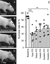
Effect of CST lesion and spontaneous recovery of skilled forelimb function on the irregular horizontal ladder. A, Uninjured animals precisely grasp each rung with all four digits placed in front of the metal bar. B–D, After unilateral CST injury, the following mistakes could be observed: animals misplaced one or more digits on the backside of the rung (B); failed to place the palm of their paw directly onto the rung (C); slipped off the rung or placed their paw between single rungs (D). E, Success rate of each animal was expressed as percentage of correct steps of all steps taken by the impaired forelimb. Pyramidotomy led to a significantly lower success rate of the impaired forelimb on the horizontal ladder (ANOVA, F = 113.16, p ≤ 0.001; n = 7). Some spontaneous improvement occurred in all animals within the next 3 weeks, but success rate remained low (Bonferroni's post hoc, p ≤ 0.01). PO, Postoperative. Data are presented as means ± SEM; single data points (black circles) represent single animals. **p ≤ 0.01; ***p ≤ 0.001. Scale bar, 1 cm.
Figure 3.
Figure 3.
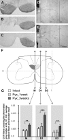
Lesion-induced growth of the intact CST across the midline and into the denervated gray matter. A, Bilateral BDA pressure injections into the forelimb motor cortex label both CST tracts. B, C, Complete interruption of one CST at the level of the caudal medulla oblongata (B) leads to interruption of BDA transport caudal to the injury (C). D, E, Representative pictures of BDA-labeled CST fibers in the contralateral gray matter of intact (D) or injured (E) animals 3 weeks after injury. F, Fibers of the intact CST were quantified by counting all intersections with lines M, D1, and D2. M was placed vertically through the midline. D1 and D2 were drawn parallel to M at one-third and two-thirds of the distance between the central canal and the lateral gray matter border. G, In intact rats (intact; n = 7), only few CST axons crossed the midline and branching was minimal. One week after injury (Pyx_1 week; n = 5), there was no change in the amount of labeled CST fibers. After 3 weeks (Pyx_3 weeks; n = 7), the number of CST collaterals projecting over the midline and branching within the denervated gray matter significantly increased (ANOVA, Bonferroni's post hoc: M, p ≤ 0.01; D1, p ≤ 0.001; D2, p ≤ 0.01). Data are presented as means ± SEM. **p ≤ 0.01; ***p ≤ 0.001. Scale bars: A–C, 200 μm; D, E, 100 μm.
Figure 4.
Figure 4.
Growth of CST fibers within lamina VII of the denervated cervical spinal cord. A, 2G13 antibodies (green) specifically label axonal growth cones as demonstrated in the developing rat neocortex (P4). Arrowheads, 2G13-positive growth cones. B, Constitutive expression of 2G13 was absent in the adult spinal cord (intermediate zone) C, Schematic spinal cord cross section with sampling area for D. D, Fibers of the intact CST (labeled with BDA; red) within the denervated gray matter. E, Terminal end of BDA labeled CST fiber. F, 2G13-positive growth cone in the denervated gray matter of fiber shown in D. G–I, Confocal analysis reveals colocalization of BDA (red) and 2G13 (green) at the terminal end of CST fibers 1 week (G, H) and 3 weeks (I) after injury demonstrating growth of CST fibers within the denervated gray matter. Scale bars: A, B, 10 μm; D, 50 μm; E–I, 2 μm.
Figure 5.
Figure 5.
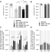
Effect of forced limb use on the recovery of skilled forelimb function and growth of the intact CST into the denervated gray matter. A, After lesion, animals were forced to completely rely on their unimpaired or their impaired limb for either 1 or 3 weeks. One week after injury, forced nonuse (Pyx_1 week_nonuse; n = 7) led to a significantly lower success rate on the horizontal ladder compared with animals that were forced to rely on their impaired limb (Pyx_1 week_use; n = 7; Bonferroni's post hoc, p ≤ 0.01). After 3 weeks, forced limb use led to full behavioral recovery back to preinjury baseline levels (Pyx_3 weeks_use; n = 8); animals that could not use their impaired side stayed significantly impaired (Pyx_3 weeks_nonuse; n = 8; p ≤ 0.001). B, Forced nonuse or forced use alone did not influence locomotor performance in sham-operated animals (sham_nonuse, n = 6; sham_use, n = 9). C, Growth and arborization of CST fibers was analyzed by counting all intersections with lines M, D1, and D2 in the denervated spinal cord; 1 week after injury, there was no difference in the number of CST fibers in animals that did use their impaired limb and animals that could not use their impaired side. Three weeks after injury, denervation led to significant growth of CST fibers across the midline in both groups (M, p ≤ 0.01). Forced limb use further increased CST arborization (D1, p ≤ 0.01; D2, p ≤ 0.05; ANOVA followed by Bonferroni's post hoc). D, Forced nonuse or forced use alone did not influence growth and arborization of CST fibers in the contralateral, manipulated gray matter (sham_nonuse, n = 6; sham_use, n = 9; ANOVA, p < 0.05). Data are presented as mean ± SEM. *p ≤ 0.05; **p ≤ 0.01; ***p ≤ 0.001.
Figure 6.
Figure 6.
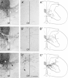
Representative pictures and camera lucida reconstructions of BDA-labeled CST fibers growing toward the contralateral denervated gray matter. Camera lucida reconstructions were made of three consecutive cross sections (50 μm; cervical segment C6). BDA-labeled fibers are depicted in black. A–A″, Intact rat. Few fibers of the intact CST cross the midline at the cervical level (M) to innervate the contralateral gray matter (D1). B–B″, Pyramidotomy, 3 weeks forced nonuse: Denervation leads to increased growth of fibers over the midline into the contralateral side as well as arborization within the denervated gray matter. C–C″, Pyramidotomy, 3 weeks forced use: Lesion induced increase of midline-crossing fibers is similar to animals that did not use their impaired limb (compare B, C). Forced limb use leads to a significant increase in arborization of fibers within the denervated gray matter. Growth and sprouting of ipsilateral ventral projections contribute to the increased fiber density. CST fibers also extended arbors deeper into dorsal or ventral laminas. Arrowheads, Midline-crossing fiber, ipsilateral ventral projection. Scale bar, 100 μm.
Figure 7.
Figure 7.
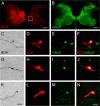
BDA-positive boutons colocalize with vGlut1, a presynaptic marker for excitatory synapses. C, G, K, Bouton-like structures were observed along the length and at the tip of BDA-labeled CST collaterals (DAB staining). A, Three weeks after injury and forced limb use, CST fibers were labeled with a fluorescent marker for BDA. Fibers were found at high densities within intermediate zone of the denervated spinal cord. Square, Sampling area. D, H, L, BDA efficiently filled collaterals of corticospinal axons up to their presumed terminal boutons. B, Intact animals show vGlut1 immunoreactivity throughout the gray matter, being strongest in superficial laminas and weaker in intermediate and ventral laminas. E, I, M, Medium-sized to large vGlut1-positive varicosities were found within the denervated gray matter. F, J, N, Confocal microscopy revealed consistent colocalization of BDA and vGlut1. Scale bars: A, B, 200 μm; C, G, K, 5 μm; D–F, H–J, L–N, 2 μm.
Figure 8.
Figure 8.
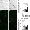
Effect of lesion and forced limb use on the number of synaptic varicosities in the denervated gray matter. C, One week after injury, the number of varicosities did not increase in animals that did not use their impaired limb (Pyx_1 week_nonuse; n = 7) or animals that completely relied on their impaired limb (Pyx_1 week_use; n = 7) compared with intact rats (intact; n = 7). After 3 weeks, forced limb use (Pyx_3 weeks_use; n = 8) led to a significant increase of boutons per fiber compared with intact, injured but freely moving, unrestricted animals (Pyx_3 weeks_free use) or animals that could not use their impaired limb (Pyx_3 weeks_nonuse; n = 8) (ANOVA, Bonferroni's post hoc, p ≤ 0.001). A, B, Representative pictures of BDA-labeled CST fibers with synaptic boutons in the contralateral gray matter are shown for animals that were restricted from using their impaired limb (A) and animals forced to use their impaired limb (B) 3 weeks after injury. A, Inset, Schematic drawings of spinal cord cross section with sampling area. Arrowheads, Boutons along CST collaterals. D–H, vGlut1-positive varicosities were counted in the intact and the denervated gray matter (n = 4/group; sampling area A; C6). Denervation led to a significant decrease of vGlut1-positive varicosities at intermediate zone in both groups 1 week after injury (p ≤ 0.01). Three weeks after injury, this decrease was still persistent (p ≤ 0.01) and there was no difference between groups (ANOVA, p > 0.05). Representative pictures of vGlut1-positive varicosities in the contralateral gray matter are shown for intact (D) and lesioned animals 1 week [forced use (E)] or 3 weeks after injury [forced nonuse (F); forced use (G)]. Data are presented as mean ± SEM. **p ≤ 0.01; ***p ≤ 0.001. Arrowheads, vGlut1-positive varicosities. Scale bars: A, B, 20 μm; D–G, 10 μm.
Figure 9.
Figure 9.
Microdissection and gene chip analysis of the denervated ventral horn. A, Schematic drawing of cross section with the extracted area. B, Spinal cord cross section with extracted ventral horn (250 μm; fresh tissue). C, Differentially regulated genes (forced limb use compared with forced nonuse) were sorted in 17 categories, and the number of regulated genes and percentages are shown for sham-operated animals as well as lesioned animals forced to rely on their impaired limb or animals that could not use their impaired limb in a table and a graphical representation. Note the high proportion of genes involved in growth, cytoskeletal reorganization, adhesion, and synapse formation in response to forced limb use in the denervated gray matter. Scale bar, 100 μm.

Similar articles

Cited by

References

    1. Adams JC. Biotin amplification of biotin and horseradish peroxidase signals in histochemical stains. J Histochem Cytochem. 1992;40:1457–1463. - PubMed
    1. Barbeau H, Rossignol S. Enhancement of locomotor recovery following spinal cord injury. Curr Opin Neurol. 1994;7:517–524. - PubMed
    1. Bareyre FM, Schwab ME. Inflammation, degeneration and regeneration in the injured spinal cord: insights from DNA microarrays. Trends Neurosci. 2003;26:555–563. - PubMed
    1. Bareyre FM, Haudenschild B, Schwab ME. Long-lasting sprouting and gene expression changes induced by the monoclonal antibody IN-1 in the adult spinal cord. J Neurosci. 2002;22:7097–7110. - PMC - PubMed
    1. Bareyre FM, Kerschensteiner M, Raineteau O, Mettenleiter TC, Weinmann O, Schwab ME. The injured spinal cord spontaneously forms a new intraspinal circuit in adult rats. Nat Neurosci. 2004;7:269–277. - PubMed

Publication types

MeSH terms

LinkOut - more resources