. 2008 Sep 17;28(38):9451-62.
doi: 10.1523/JNEUROSCI.2674-08.2008.

Systemic inflammation alters the kinetics of cerebrovascular tight junction disruption after experimental stroke in mice

Affiliations

Systemic inflammation alters the kinetics of cerebrovascular tight junction disruption after experimental stroke in mice

Barry W McColl et al. J Neurosci. .

Abstract

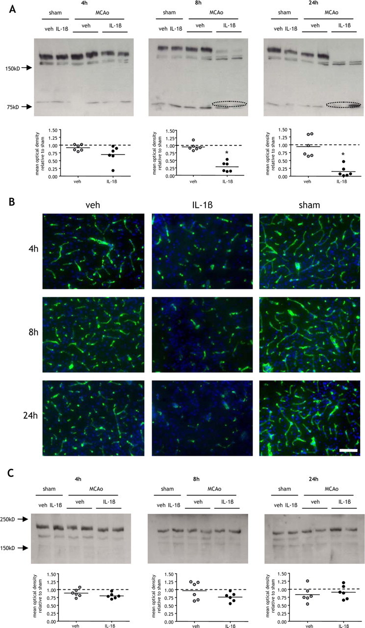
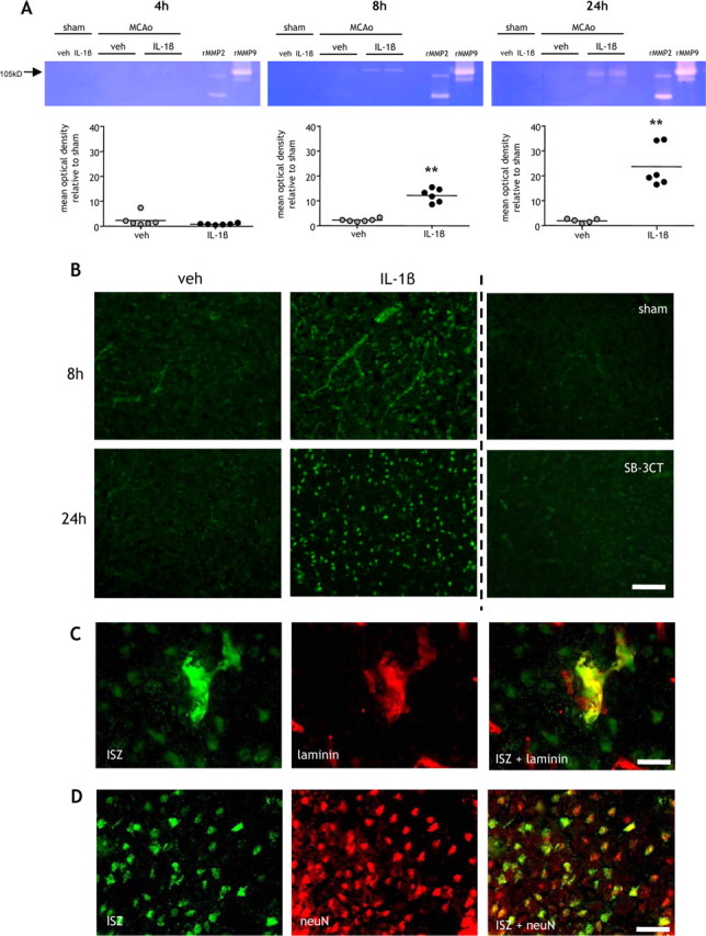
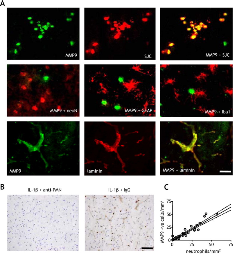
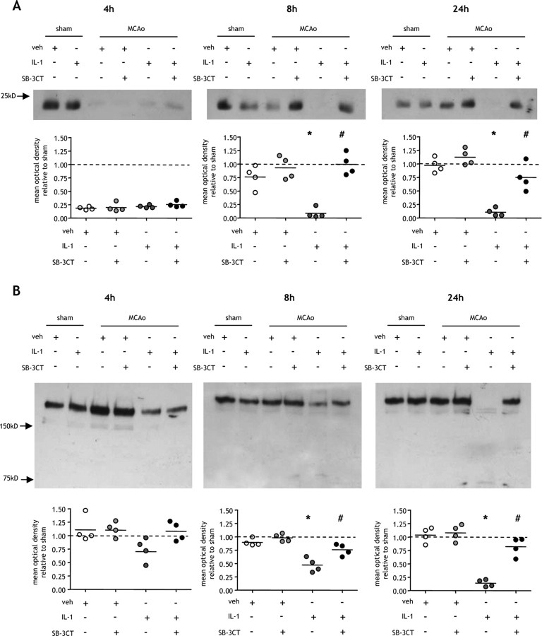
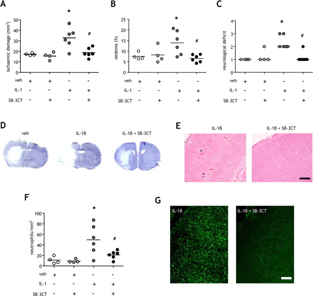
Systemic inflammatory events, such as infection, increase the risk of stroke and are associated with worse outcome, but the mediators of this clinically important effect are unknown. Our aim here was to elucidate mechanisms contributing to the detrimental effects of systemic inflammation on mild ischemic brain injury in mice. Systemic inflammation was induced in mice by peripheral interleukin-1beta (IL-1beta) challenge and focal cerebral ischemia by transient middle cerebral artery occlusion (MCAo). Systemic inflammation caused an alteration in the kinetics of blood-brain barrier (BBB) disruption through conversion of a transient to a sustained disruption of the tight junction protein, claudin-5, and also markedly exacerbated disruption to the cerebrovascular basal lamina protein, collagen-IV. These alterations were associated with a systemic inflammation-induced increase in neurovascular gelatinolytic activity that was mediated by a fivefold increase in neutrophil-derived matrix metalloproteinase-9 (MMP-9) in the brains of IL-1beta-challenged mice after MCAo. Specific inhibition of MMP-9 abrogated the effects of systemic inflammation on the sustained but not the acute disruption of claudin-5, which was associated with phosphorylation of cerebrovascular myosin light chain. MMP-9 inhibition also attenuated the deleterious impact of systemic inflammation on brain damage, edema, neurological deficit, and incidence of hemorrhagic transformation. These data indicate that a transformation from transient to sustained BBB disruption caused by enhanced neutrophil-derived neurovascular MMP-9 activity is a critical mechanism underlying the exacerbation of ischemic brain injury by systemic inflammation. These mechanisms may contribute to the poor clinical outcome in stroke patients presenting with antecedent infection.


Figures

Figure 1.
Figure 1.
Systemic inflammation causes sustained claudin-5 disruption after MCAo. Systemic inflammation was induced by intraperitoneal IL-1β injection, and the effects on the BBB tight junction proteins, claudin-5 and occludin, were assessed in cortical tissue ipsilateral to MCAo by immunoblot and immunofluorescence at indicated times after MCAo. A, MCAo induced a 50% reduction in claudin-5 levels in both vehicle (veh)- and IL-1β-treated mice compared with sham-operated mice after 4 h reperfusion. This disruption to claudin-5 resolved in vehicle-treated mice by 8 h after MCAo and was maintained at 24 h in which claudin-5 levels were similar to sham-operated mice. There was a sustained disruption to claudin-5 in IL-1β-treated mice 8 and 24 h after MCAo resulting in a 70% reduction compared with sham-operated mice at 24 h. The sustained loss of claudin-5 in IL-1β-challenged mice resulted in a significant reduction compared with vehicle-treated mice 8 and 24 h after MCAo. B, Claudin-5 immunofluorescence (green channel) with DAPI counterstain (blue channel) demonstrated dense networks of cerebrovascular immunoreactivity in sham-operated mice. Marked reductions in immunoreactivity were evident 4 h after MCAO in vehicle-treated mice and 4, 8, and 24 h after MCAo in IL-1β-challenged mice, consistent with the immunoblot data. C, Confocal immunofluorescence revealed the disruption by systemic inflammation to the highly organized arrangement of claudin-5 in the interendothelial tight junctions of cortical blood vessels in the ipsilateral hemisphere 24 h after MCAo. D, There were no significant effects of systemic inflammation or MCAo on occludin levels. All immunofluorescence images are from the cortex ipsilateral to MCAo. *p < 0.05 versus vehicle; Student's t test. Scale bars: B, 100 μm; C, 10 μm.
Figure 2.
Figure 2.
Systemic inflammation aggravates cerebrovascular collagen-IV disruption after MCAo. Systemic inflammation was induced by intraperitoneal IL-1β injection and the effects on the cerebrovascular basement lamina proteins, collagen-IV, and laminin, were assessed in cortical tissue ipsilateral to MCAo by immunoblot and immunofluorescence at indicated times after MCAo. A, Collagen-IV levels in vehicle (veh)-treated mice were similar in sham-operated mice and after MCAo at all time points examined. IL-1β challenge caused a progressive decline in collagen-IV as reperfusion progressed and resulted in an 80% reduction compared with sham levels 24 h after MCAo. There was a significant reduction in collagen-IV levels in IL-1β-challenged mice compared with vehicle-treated mice 8 and 24 h after MCAo, and a putative cleavage collagen-IV cleavage product (circled) was evident in IL-1β-challenged mice at these time points. B, Collagen-IV immunofluorescence (green channel) with DAPI counterstain (blue channel) demonstrated extensive and dense networks of cerebrovascular immunoreactivity in sham-operated mice and a similar pattern was evident in vehicle-treated mice after MCAo. IL-1β challenge induced a marked reduction in collagen-IV immunoreactivity 4, 8, and 24 h after MCAo. C, Systemic inflammation or MCAo did not significantly affect laminin levels. All immunofluorescence images are from the cortex ipsilateral to MCAo. *p < 0.05 versus vehicle; Student's t test. Scale bar, 50 μm.
Figure 3.
Figure 3.
Systemic inflammation exacerbates neurovascular gelatinolytic activity. A, B, Systemic inflammation was induced by intraperitoneal IL-1β injection and the extent of gelatinolytic activity determined by gel zymography in purified cortical brain samples (A) and by in situ zymography on brain sections at indicated times after MCAo (B). A, Gelatinolytic activity was minimal 4 h after MCAo, and there was no difference between vehicle (veh)- and IL-1β-treated mice. Clear gelatinolytic bands corresponding to pro-MMP-9 (105 kDa) and active MMP-9 (97 kDa) were evident 8 and 24 h after MCAo in vehicle- and IL-1β-treated mice but not after sham occlusion. The extent of gelatinolytic activity (105 kDa) was significantly greater in IL-1β-treated mice 8 and 24 h after MCAo. There was no detectable gelatinolytic activity corresponding to MMP-2 (72 kDa). B, In situ zymography demonstrated that increased gelatinolytic activity in IL-1β-challenged mice was primarily localized to the cerebral vasculature and neurons 8 and 24 h after MCAo. In situ gelatinolytic activity was minimal in sham-operated mice and attenuated by coincubation with the MMP-9 inhibitor, SB-3CT. C, Immunofluorescence was combined with in situ zymography to confirm the localization of gelatinolytic activity. D, Gelatinolytic activity colocalized with the vascular basement membrane protein, laminin, and the neuronal marker neuN. **p < 0.01 versus vehicle; Student's t test. Scale bars: B, 100 μm; C, 15 μm; D, 25 μm.
Figure 4.
Figure 4.
Systemic inflammation increases cerebral MMP-9 immunoreactivity after MCAo. Systemic inflammation was induced by intraperitoneal IL-1β injection and MMP-9 immunostaining assessed at indicated times after MCAo. A–C, MMP-9 immunostaining was evident in cells adherent to cortical penetrating vessels (A) and microvessels (B) and in cells throughout the parenchyma (C) in the hemisphere ipsilateral to MCAo. There was minimal immunoreactivity in the contralateral hemisphere and in sham-operated mice. D, Representative brain sections illustrate the marked increase in MMP-9-immunoreactive cells in the cortex of IL-1β-treated mice 8 and 24 h after MCAo. E, Quantification of cellular MMP-9 immunoreactivity. Systemic inflammation significantly increased the number of MMP-9+ cells in the cortex 4, 8, and 24 h after MCAo and in the striatum 8 h after MCAo. *p < 0.05, **p < 0.01 versus vehicle (veh); Student's t test. Scale bars: (in C) A–C, 25 μm; D, 100 μm.
Figure 5.
Figure 5.
Infiltrating neutrophils are the primary source of MMP-9 in the ischemic brain. The cellular localization of MMP-9 immunoreactivity in the ischemic brain was examined by double-labeling immunofluorescence. A, There was strong and extensive colocalization between MMP-9 and the specific neutrophil marker, SJC, but not with neuronal (neuN), astrocytic (GFAP), or microglial (Iba1) markers. There was also strong colocalization of MMP-9 with the vascular basement membrane protein, laminin. B, Depletion of neutrophils using an anti-PMN antibody abolished the cellular and vascular MMP-9 immunoreactivity in the brain, but there was no effect with an isotype control antibody. C, There was a strong correlation between the number of MMP+ cells and the number of neutrophils in the cortex of vehicle- and IL-1β-treated mice. R2 = 0.91; p < 0.01, Pearson correlation. All double-labeling immunofluorescence images are from the cortex ipsilateral to MCAo of IL-1β-treated mice 24 h after MCAo. Scale bars: A, 25 μm; B, 100 μm.
Figure 6.
Figure 6.
Sustained aggravation of postischemic BBB disruption caused by systemic inflammation is dependent on MMP-9. A, B, Systemic inflammation was induced by intraperitoneal IL-1β injection, and the effects of MMP-9 inhibition using the specific inhibitor, SB-3CT, on the BBB tight junction protein, claudin-5 (A), and the cerebrovascular basal lamina protein, collagen-IV (B), were assessed in cortical tissue ipsilateral to MCAo by immunoblot at indicated times after MCAo. A, MCAo induced a 75% reduction in claudin-5 levels after 4 h reperfusion in vehicle (veh)- and IL-1β-treated mice, and there was no effect of MMP-9 inhibition at this time point. Claudin-5 levels were restored to sham occlusion levels 8 and 24 h after MCAo, and there was no significant effect of MMP-9 inhibition. Peripheral inflammatory challenge caused a sustained reduction in claudin-5 8 and 24 h after MCAo, and these effects were significantly attenuated by inhibition of MMP-9. B, Collagen-IV levels were similar after sham occlusion and MCAo in vehicle-treated mice at all time points examined. Peripheral inflammatory challenge induced a significant reduction in collagen-IV 8 and 24 h after MCAo, and these effects were significantly attenuated by inhibition of MMP-9. *p < 0.05 versus vehicle, #p < 0.05 versus IL-1β, Kruskal–Wallis test followed by Dunn's multiple-comparison test.
Figure 7.
Figure 7.
Inhibition of MMP-9 ameliorates the exacerbation of ischemic brain damage by systemic inflammation. Systemic inflammation was induced by intraperitoneal IL-1β injection, and the effects of MMP-9 inhibition using the specific inhibitor, SB-3CT, on the extent of ischemic brain damage, brain edema, and the severity of neurological deficit were determined 24 h after MCAo. A–C, Systemic inflammation significantly exacerbated the volume of ischemic damage (A), the extent of brain edema (B), and the neurological deficit (C). Inhibition of MMP-9 significantly attenuated the deleterious effects of systemic inflammation on brain damage (A), edema (B), and neurological deficit (C). D, Representative cresyl violet-stained brain sections illustrate the exacerbation of brain damage by systemic inflammation and the attenuation of this effect by SB-3CT. E, SB-3CT prevented the hemorrhagic transformation (arrowheads) caused by systemic inflammation. F, G, Inhibition of MMP-9 also attenuated the potentiation of neutrophil infiltration (F) and gelatinolytic activity (G) caused by peripheral inflammatory challenge. A, B, F: *p < 0.05 versus vehicle (veh), #p < 0.05 versus IL-1β, one-way ANOVA followed by Student's t test with Bonferroni correction; C: *p < 0.05 versus vehicle, #p = 0.05 versus IL-1β, generalized Fisher's exact test with Bonferroni correction. Scale bars, 100 μm.
Figure 8.
Figure 8.
Temporal involvement of MMP-9 in BBB disruption underlying the exacerbation of ischemic brain damage by systemic inflammation. Our data indicate two phases of BBB disruption based on the involvement of MMP-9. The first phase is MMP-9-independent and also occurs independently of systemic inflammation suggesting a local, CNS-driven mechanism such as the cytokine/ROS-induced phosphorylation of endothelial myosin light chain. This phase of BBB disruption is reversible when there is no additional peripheral inflammatory stimulus. Systemic inflammation causes a secondary MMP-9-dependent phase of disruption that is systemically driven (likely by neutrophils) and compounds the initial disturbance and results in sustained and irreversible BBB dysfunction. The deleterious effects of systemic inflammation on ischemic brain damage highlight the important position of the BBB as a point of convergence for CNS-driven and systemically driven inflammatory processes. veh, Vehicle.

References

    1. Afonso PV, Ozden S, Prevost MC, Schmitt C, Seilhean D, Weksler B, Couraud PO, Gessain A, Romero IA, Ceccaldi PE. Human blood-brain barrier disruption by retroviral-infected lymphocytes: role of myosin light chain kinase in endothelial tight-junction disorganization. J Immunol. 2007;179:2576–2583. - PubMed
    1. Alvarez JI, Teale JM. Evidence for differential changes of junctional complex proteins in murine neurocysticercosis dependent upon CNS vasculature. Brain Res. 2007;1169:98–111. - PMC - PubMed
    1. Asahi M, Wang X, Mori T, Sumii T, Jung JC, Moskowitz MA, Fini ME, Lo EH. Effects of matrix metalloproteinase-9 gene knock-out on the proteolysis of blood-brain barrier and white matter components after cerebral ischemia. J Neurosci. 2001;21:7724–7732. - PMC - PubMed
    1. Bederson JB, Pitts LH, Tsuji M, Nishimura MC, Davis RL, Bartkowski H. Rat middle cerebral artery occlusion: evaluation of the model and development of a neurologic examination. Stroke. 1986;17:472–476. - PubMed
    1. Belayev L, Busto R, Zhao W, Ginsberg MD. Quantitative evaluation of blood-brain barrier permeability following middle cerebral artery occlusion in rats. Brain Res. 1996;739:88–96. - PubMed

Publication types

MeSH terms