. 2008 Sep 17;28(38):9585-97.
doi: 10.1523/JNEUROSCI.5698-07.2008.

Decreased striatal dopamine release underlies increased expression of long-term synaptic potentiation at corticostriatal synapses 24 h after 3-nitropropionic-acid-induced chemical hypoxia

Affiliations

Decreased striatal dopamine release underlies increased expression of long-term synaptic potentiation at corticostriatal synapses 24 h after 3-nitropropionic-acid-induced chemical hypoxia

Garnik Akopian et al. J Neurosci. .

Abstract

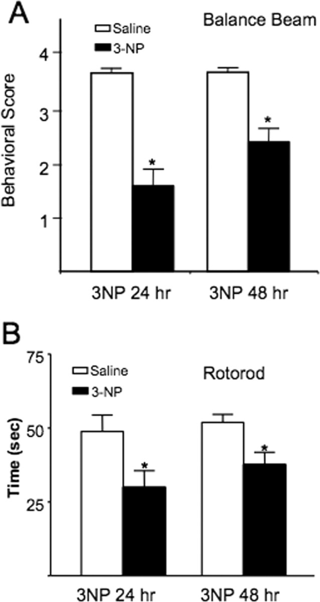
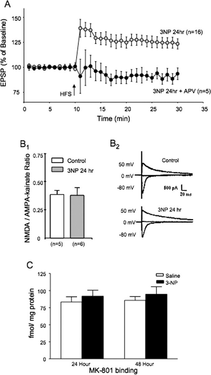
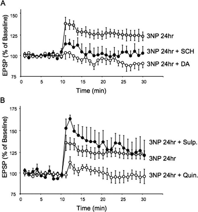
The striatum is particularly sensitive to the irreversible inhibitor of succinate dehydrogenase 3-nitropropionic acid (3-NP). In the present study, we examined early changes in behavior and dopamine and glutamate synaptic physiology created by a single systemic injection of 3-NP in Fischer 344 rats. Hindlimb dystonia was seen 2 h after 3-NP injections, and rats performed poorly on balance beam and rotarod motor tests 24 h later. Systemic 3-NP increased NMDA receptor-dependent long-term potentiation (LTP) at corticostriatal synapses over the same time period. The 3-NP-induced corticostriatal LTP was not attributable to increased NMDA receptor number or function, because 3-NP did not change MK-801 [(+)-5-methyl-10,11-dihydro-5H-dibenzo[a,d]cyclohepten-5,10-imine] binding or NMDA/AMPA receptor current ratios. The LTP seen 24 h after 3-NP was D(1) receptor dependent and reversed by exogenous addition of dopamine or a D(2) receptor agonist to brain slices. HPLC and fast-scan cyclic voltammetry revealed a decrease in dopamine content and release in rats injected 24 h earlier with 3-NP, and much like the enhanced LTP, dopamine changes were reversed by 48 h. Tyrosine hydroxylase expression was not changed, and there was no evidence of striatal cell loss at 24-48 h after 3-NP exposure. Sprague Dawley rats showed similar physiological responses to systemic 3-NP, albeit with reduced sensitivity. Thus, 3-NP causes significant changes in motor behavior marked by parallel changes in striatal dopamine release and corticostriatal synaptic plasticity.


Figures

Figure 1.
Figure 1.
Systemic 3-NP causes impairment in balance beam and rotarod performance during the first 48 h in rats. A, Rats were ranked according to a five-point scale on their ability to navigate a balance beam according to the methods outlined by Friedemann and Gerhardt (1992). Rats showed impairment at both 24 and 48 h after systemic injection with 3-NP (*p < 0.01). B, Rats were tested on a slowly accelerating rotarod test and both the 24 and 48 h after 3-NP rats showed impairment relative to their performance before being exposed to 3-NP (*p < 0.01).
Figure 2.
Figure 2.
Systemic exposure to 3-NP increases LTP expression at dorsomedial corticostriatal synapses. A, Example traces of EPSPs selected from indicated time points for each experimental group. B, Plots of tetanus-induced long-term synaptic plasticity at dorsomedial corticostriatal synapses are shown. 3-NP injected intraperitoneally 24 h earlier increased the expression of LTP induced in vitro. Control rats were injected with saline and examined 24 h later. Long-term plasticity switched to LTD by 48 h after the 3-NP injection. EPSPs were measured using sharp electrode intracellular recordings. C, Bar graphs showing distribution of cells expressing tetanus-induced potentiation (>100%) and depression (<100%) for STP and LTP under each experimental condition (control, 24 and 48 h after 3-NP).
Figure 3.
Figure 3.
LTP produced by systemic 3-NP is NMDA receptor dependent. A, Plots of tetanus-induced long-term synaptic plasticity in rats injected 24 h earlier with 3-NP. The 3-NP group is the same as that shown in Figure 1, where brain slices were perfused with normal aCSF. Perfusion of brain slices taken from rats injected 24 h earlier with 3-NP with the NMDA receptor antagonist APV blocked the induction of LTP seen in normal aCSF. B, The NMDA/AMPA-kainate synaptic current ratio does not change at corticostriatal synapses from rats injected 24 h earlier with 3-NP. B1, Bar graph of NMDA/AMPA-kainate current synaptic ratio from saline and 3-NP-injected rats. B2, Examples of corticostriatal synaptic currents evoked at the indicated holding potentials using single electrode switch clamp methods. C, MK-801 binding to NMDA receptors from the dorsomedial striatum does not change 24 h after systemic injection with 3-NP. Bar graphs illustrate MK-801 binding in saline-injected and 3-NP-injected rats (24 and 48 h after 3-NP).
Figure 4.
Figure 4.
LTP produced by systemic injection of 3-NP 24 h earlier requires dopamine activation of D1 receptors. A, LTP produced by systemic injection of 3-NP is eliminated by addition of the D1 receptor antagonist SCH 23990 or dopamine. Plots of tetanus-induced plasticity in brain slices taken from rats injected 24 h earlier with 3-NP. Brain slices were bathed in either aCSF, aCSF + SCH 23390 (10 μm), or aCSF + 30 μm dopamine. B, LTP produced by systemic injection of 3-NP 24 h earlier is slightly enhanced by block of D2 receptors with l-sulpiride (Sulp) (1 μm) and completely eliminated by activation of D2 receptors with the D2 receptor agonist quinpirole (Quin) (10 μm).
Figure 5.
Figure 5.
Systemic 3-NP causes a reduction in dopamine content in the dorsomedial striatum 24 h after the injection. A, Bar graphs of dopamine content, DOPAC, and HVA obtained from the dorsomedial striatum of rats injected with 3-NP (16.5 mg/kg) 24 and 48 h earlier. The single injection of 3-NP caused a significant reduction in dopamine content 24 h after the injection, but this change reversed by 48 h after the injection. B, Injection of 3-NP for 2 consecutive days causes significant changes in dorsomedial striatal dopamine, DOPAC, and HVA. The double injection protocol was 15 mg/kg on day 1 and 10 mg/kg on day 2, and the rat was killed 3 h later for analysis of dopamine and its metabolites in total striatal tissue. Dopamine content was dramatically reduced, and this change was associated with large changes in DOPAC and HVA. Note the change in scale for the DOPAC measurement for the double injection protocol. The single injection data from the dorsomedial striatum shown in A are illustrated for comparison. All values are normalized to saline-injected control rats. *p < 0.05; **p < 0.03.
Figure 6.
Figure 6.
Systemic injection of 3-NP creates regionally dependent decreases in dopamine release in the striatum. The amount of dopamine release was determined at 5 regions within the striatum (at approximately bregma level 1.00–0.80) including (1) midstriatum, (2) dorsomedial, (3) dorsal, (4) dorsolateral, and (5) ventrolateral in representative mice from all four groups (see brain slice inset). The bar graph illustrates peak dopamine released by a single intrastriatal stimulus (Calibration: 200 μA, 0.1 ms) applied to striatal brain slices at each of the above indicated regions for saline-injected rats, rats injected 24 h earlier with 3-NP, and rats injected 48 h earlier with 3-NP. Inset, Voltammogram response for dopamine release. Fifty traces are shown at a sampling rate of every 100 ms (10 Hz) after delivery of a single 0.1 ms, 200 μA intrastriatal stimulus (*p < 0.05 for comparison of saline control vs 24 after 3-NP injection).
Figure 7.
Figure 7.
Staining of striatal tissue for tyrosine hydroxylase protein or Nissl substance. A–L, Tissue sections from the midstriatum of rats treated with either saline or 3-NP (collected at 1 or 2 d after lesioning) were immunostained for either TH protein (A–F) or Nissl substance (G–L). The top panels show sections at low magnification of the entire striatum in coronal sections, whereas the bottom panels show a higher magnification (20×) of the dorsal region of the striatum. Scale bars: (top) 1.7 mm; (bottom) 100 μm. Relative optical density of the dorsomedial striatum (graph below TH panels) showed no evidence of loss of TH immunostaining, and cell counting (graph below Nissl panels) showed no reduced number of Nissl-stained striatal neurons.
Figure 8.
Figure 8.
Sprague Dawley rats show reduced sensitivity to systemic 3-NP. A, Plots of tetanus-induced long-term synaptic plasticity at dorsomedial corticostriatal synapses are shown. No difference was found in the tetanus-induced plasticity between slices taken from saline-injected Sprague Dawley rats and Sprague Dawley rats injected 24 h earlier with 25–35 mg/kg 3-NP. A trend toward reduced LTD was seen in slices taken from rats injected once a day for 3 d with 3-NP (15, 10, and 10 mg/kg). B, Bar graphs showing distribution of cells expressing tetanus-induced potentiation (>100%) and depression (<100%) for STP and LTP under each experimental condition (saline, single injection, and 3× injection). C, Systemic injection of 3-NP creates regionally dependent decreases in dopamine release in the striatum. The amount of dopamine release was determined at 5 regions within the striatum including (1) midstriatum, (2) dorsomedial, (3) dorsal, (4) dorsolateral, and (5) ventrolateral (inset). Bar graph illustrates peak dopamine released by a single intrastriatal stimulus (200 μA, 0.1 ms) applied to striatal brain slices at each of the above indicated regions for saline-injected rats, rats injected 24 h earlier with 3-NP (25–35 mg/kg 3-NP), and rats injected once a day for 3 d with 3-NP (15, 10, and 10 mg/kg) and killed 24 h after the last injection. No differences were observed between saline-injected and single injected (25–35 mg/kg 3-NP) Sprague Dawley rats. A significant reduction in dopamine release was seen in rats injected once a day for 3 d with 3-NP across all 5 sampling sites (repeated-measures ANOVA; p < 0.03). Post hoc analysis showed reduced release at sites 1, 3, 4 (*p < 0.01) and 2 (**p < 0.02).

References

    1. Akopian G, Walsh JP. Reduced expression of short- and long-term facilitation at aged corticostriatal synapses. Synapse. 2006;60:223–238. - PubMed
    1. Akopian G, Musleh W, Smith R, Walsh JP. Functional state of presynaptic terminals influences the expression of short- and long-term plasticity at corticostriatal synapses. Synapse. 2000;38:271–280. - PubMed
    1. Ariano MA, Monsma FJ, Jr, Barton AC, Kang HC, Haugland RP, Sibley DR. Direct visualization and cellular localization of D1 and D2 dopamine receptors in rat forebrain by use of fluorescent ligands. Proc Natl Acad Sci U S A. 1989;86:8570–8574. - PMC - PubMed
    1. Aspey BS, Cohen S, Patel Y, Terruli M, Harrison MJ. Middle cerebral artery occlusion in the rat: consistent protocol for a model of stroke. Neuropathol Appl Neurobiol. 1998;24:487–497. - PubMed
    1. Aspey BS, Taylor FL, Terruli M, Harrison MJ. Temporary middle cerebral artery occlusion in the rat: consistent protocol for a model of stroke and reperfusion. Neuropathol Appl Neurobiol. 2000;26:232–242. - PubMed

Publication types

MeSH terms

LinkOut - more resources