

doi: 10.1128/JCM.00482-08.
Epub 2008 Sep 17.
Development of natural culture media for rapid induction of Fonsecaea pedrosoi sclerotic cells in vitro
Affiliations
- PMID: 18799695
- PMCID: PMC2576606
- DOI: 10.1128/JCM.00482-08
Item in Clipboard
Development of natural culture media for rapid induction of Fonsecaea pedrosoi sclerotic cells in vitro
J Clin Microbiol.
2008 Nov.
Abstract
Fonsecaea pedrosoi is the main agent of chromoblastomycosis, a skin disease presenting verrucous lesions, in which round, thick-walled sclerotic cells are found. In vitro induction of sclerotic cells is time-consuming (20 to 45 days) and temperature dependent. We present two new natural media that reduce the sclerotic-cell induction time to only 2 days.
Figures
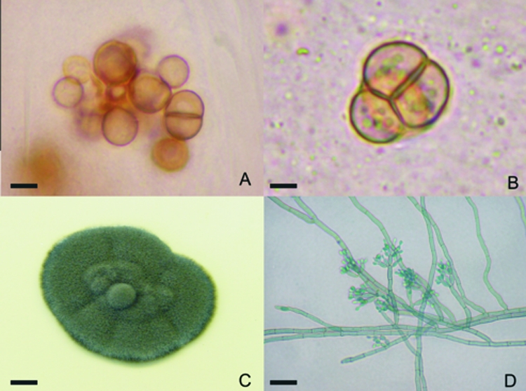
Fonsecaea pedrosoi sclerotic cells were obtained after scraping of chromoblastomycosis lesions. The cells gave rise to hyphae and conidia after in vitro culture. (A and B) Skin scrapings were collected and analyzed after clarification with 20% KOH, revealing well-defined, septated, sclerotic cells. (C) Greenish-black colonies grew from culture of this material on Mycosel. (D) Characteristic dematiaceous hyphae originating terminal cylindrical conidiophores with small subhyaline conidia were observed upon microculture. Bars, 4 μm (A), 3 μm (B), 0.5 cm (C), and 10 μm (D).
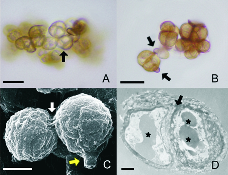
The morphological characteristics of sclerotic cells induced in either of the two natural media are similar. (A and B) Sclerotic cells obtained after culture of conidia in Theobroma grandiflorum or Bactris gasipaes natural media were analyzed by optical microscopy with no special staining. (A) Brownish, multiseptated cells (arrow) were evident. (B) Remnants of conidia adhering to the newly formed sclerotic cells (arrows) were observed. (C) SEM of aggregates of sclerotic cells demonstrates septated division (white arrow) and remnants of conidia (yellow arrow). (D) TEM of a sclerotic cell shows typical thick-walled septation (arrow) and vesicles containing electron-dense material (asterisks). Data are representative of one of three independent experiments, which were performed in triplicate. Bars, 10 μm (A and B), 5 μm (C), and 1 μm (D).
References
-
- Alviano, D. S., L. F. Kneipp, A. H. Lopes, L. R. Travassos, J. R. Meyer-Fernandes, M. L. Rodrigues, and C. S. Alviano. 2003. Differentiation of Fonsecaea pedrosoi mycelial forms into sclerotic cells is induced by platelet-activating factor. Res. Microbiol. 154689-695. - PubMed
-
- Cermeño-Vivas, J. R., and J. M. Torres Rodriguez. 2001. In vitro susceptibility of dematiaceous fungi to ten antifungal drugs using an agar diffusion test. Rev. Iberoam. Micol. 18113-117. (In Spanish.) - PubMed
-
- da Silva, J. P., D. S. Alviano, C. S. Alviano, W. de Souza, L. R. Travassos, J. A. Diniz, and S. Rozental. 2002. Comparison of Fonsecaea pedrosoi sclerotic cells obtained in vivo and in vitro: ultrastructure and antigenicity. FEMS Immunol. Med. Microbiol. 3363-69. - PubMed
-
- da Silva, J. P., M. B. da Silva, U. I. Salgado, J. A. Diniz, S. Rozental, and C. G. Salgado. 2007. Phagocytosis of Fonsecaea pedrosoi conidia, but not sclerotic cells caused by Langerhans cells, inhibits CD40 and B7-2 expression. FEMS Immunol. Med. Microbiol. 50104-111. - PubMed
-
- De Hoog, G. S., J. Guarro, J. Gené, and M. J. Figueras. 2000. Atlas of clinical fungi. Centraalbureau voor Schimmelcultures and Universitat Rovira i Virgili, Utrecht, The Netherlands.
Publication types
MeSH terms
Substances
LinkOut - more resources
Full Text Sources
